Explore
 Validate
 Learn
Learn Western blot
 Immunocytochemistry
 Immunoprecipitation
Antibody data
- Antibody Data
- Antigen structure
- References [9]
- Comments [0]
- Validations
- Immunocytochemistry [4]
- Immunohistochemistry [2]
- Other assay [3]
Submit
Validation data
Reference
Comment
Report error
- Product number
- MA3-310 - Provider product page

- Provider
- Invitrogen Antibodies
- Product name
- Estrogen Receptor alpha Monoclonal Antibody (EVG F9)
- Antibody type
- Monoclonal
- Antigen
- Synthetic peptide
- Description
- MA3-310 detects estrogen receptor alpha in human and rabbit samples. MA3-310 has been successfully used in immunohistochemistry, immunoprecipitation and Western blot procedures. By Western blot, this antibody detects a ~68 kDa protein representing estrogen receptor alpha in MCF-7 cells, human breast tumors, and rabbit vaginal tissue. In immunohistochemistry this antibody recognizes the estrogen receptor alpha in MCF-7 cells, human breast tissue, and human smooth muscle cells. The MA3-310 immunogen is a mix of synthetic peptides corresponding to residuesT(140) V R E A G P P A F Y R P N S(154) & E(247) V G M M K G G I R K D R R G G R(263) of human ER alpha.
- Reactivity
- Human, Rabbit
- Host
- Mouse
- Isotype
- IgG
- Antibody clone number
- EVG F9
- Vial size
- 100 μL
- Concentration
- Conc. Not Determined
- Storage
- -20°C, Avoid Freeze/Thaw Cycles
Submitted references Raloxifene Stimulates Estrogen Signaling to Protect Against Age- and Sex-Related Intervertebral Disc Degeneration in Mice.
Simultaneous defeat of MCF7 and MDA-MB-231 resistances by a hypericin PDT-tamoxifen hybrid therapy.
High Estradiol Differentially Affects the Expression of the Glucose Transporter Type 4 in Pelvic Floor Muscles of Rats.
Role of Estrogens in the Size of Neuronal Somata of Paravaginal Ganglia in Ovariectomized Rabbits.
Calmodulin-like protein 3 is an estrogen receptor alpha coregulator for gene expression and drug response in a SNP, estrogen, and SERM-dependent fashion.
Aromatase expression is linked to estrogenic sensitivity of periurethral muscles in female rabbits.
Oestrogen upregulates L-type Ca²⁺ channels via oestrogen-receptor- by a regional genomic mechanism in female rabbit hearts.
Immunodetection of nmt55/p54nrb isoforms in human breast cancer.
Estradiol-induced phosphorylation of serine 118 in the estrogen receptor is independent of p42/p44 mitogen-activated protein kinase.
Bhadouria N, Berman AG, Wallace JM, Holguin N
Frontiers in bioengineering and biotechnology 2022;10:924918
Frontiers in bioengineering and biotechnology 2022;10:924918
Simultaneous defeat of MCF7 and MDA-MB-231 resistances by a hypericin PDT-tamoxifen hybrid therapy.
Theodossiou TA, Ali M, Grigalavicius M, Grallert B, Dillard P, Schink KO, Olsen CE, Wälchli S, Inderberg EM, Kubin A, Peng Q, Berg K
NPJ breast cancer 2019;5:13
NPJ breast cancer 2019;5:13
High Estradiol Differentially Affects the Expression of the Glucose Transporter Type 4 in Pelvic Floor Muscles of Rats.
Carrasco-Ruiz MLÁ, Hernández-Aragón LG, Chávez-Ríos JR, Rodríguez-Antolín J, Pacheco P, Martínez-Gómez M, Cuevas-Romero E, Castelán F
International neurourology journal 2018 Sep;22(3):161-168
International neurourology journal 2018 Sep;22(3):161-168
Role of Estrogens in the Size of Neuronal Somata of Paravaginal Ganglia in Ovariectomized Rabbits.
Hernández-Aragón LG, García-Villamar V, Carrasco-Ruiz ML, Nicolás-Toledo L, Ortega A, Cuevas-Romero E, Martínez-Gómez M, Castelán F
BioMed research international 2017;2017:2089645
BioMed research international 2017;2017:2089645
Calmodulin-like protein 3 is an estrogen receptor alpha coregulator for gene expression and drug response in a SNP, estrogen, and SERM-dependent fashion.
Qin S, Ingle JN, Liu M, Yu J, Wickerham DL, Kubo M, Weinshilboum RM, Wang L
Breast cancer research : BCR 2017 Aug 18;19(1):95
Breast cancer research : BCR 2017 Aug 18;19(1):95
Aromatase expression is linked to estrogenic sensitivity of periurethral muscles in female rabbits.
de los Ángeles Carrasco-Ruiz M, García-Villamar V, López-García K, Sánchez-García O, Pacheco P, Cuevas E, Martínez-Gómez M, Castelán F
Cell biochemistry and function 2015 Jun;33(4):188-95
Cell biochemistry and function 2015 Jun;33(4):188-95
Oestrogen upregulates L-type Ca²⁺ channels via oestrogen-receptor- by a regional genomic mechanism in female rabbit hearts.
Yang X, Chen G, Papp R, Defranco DB, Zeng F, Salama G
The Journal of physiology 2012 Feb 1;590(3):493-508
The Journal of physiology 2012 Feb 1;590(3):493-508
Immunodetection of nmt55/p54nrb isoforms in human breast cancer.
Pavao M, Huang YH, Hafer LJ, Moreland RB, Traish AM
BMC cancer 2001;1:15
BMC cancer 2001;1:15
Estradiol-induced phosphorylation of serine 118 in the estrogen receptor is independent of p42/p44 mitogen-activated protein kinase.
Joel PB, Traish AM, Lannigan DA
The Journal of biological chemistry 1998 May 22;273(21):13317-23
The Journal of biological chemistry 1998 May 22;273(21):13317-23
No comments: Submit comment
Supportive validation
- Submitted by
- Invitrogen Antibodies (provider)
- Main image

- Experimental details
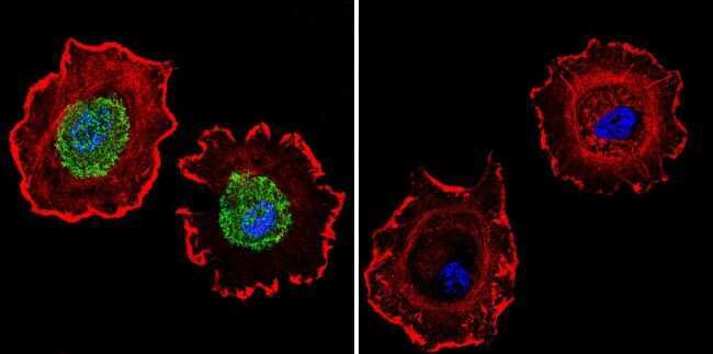
- Immunofluorescent analysis of Estrogen Receptor alpha using Anti-Estrogen Receptor alpha Monoclonal Antibody (EVG F9) (Product # MA3-310) shows staining in MCF-7 Cells. Estrogen Receptor alpha staining (green), F-Actin staining with Phalloidin (red) and nuclei with DAPI (blue) is shown. Cells were grown on chamber slides and fixed with formaldehyde prior to staining. Cells were probed without (control) or with or an antibody recognizing Estrogen Receptor alpha (Product # MA3-310) at a dilution of 1:200 over night at 4°C, washed with PBS and incubated with a DyLight-488 conjugated secondary antibody (Product # 35503, Goat Anti-Mouse). Images were taken at 60X magnification.
- Submitted by
- Invitrogen Antibodies (provider)
- Main image

- Experimental details
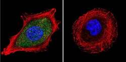
- Immunofluorescent analysis of Estrogen Receptor alpha using Anti-Estrogen Receptor alpha Monoclonal Antibody (EVG F9) (Product # MA3-310) shows staining in U251 Cells. Estrogen Receptor alpha staining (green), F-Actin staining with Phalloidin (red) and nuclei with DAPI (blue) is shown. Cells were grown on chamber slides and fixed with formaldehyde prior to staining. Cells were probed without (control) or with or an antibody recognizing Estrogen Receptor alpha (Product # MA3-310) at a dilution of 1:200 over night at 4°C, washed with PBS and incubated with a DyLight-488 conjugated secondary antibody (Product # 35503, Goat Anti-Mouse). Images were taken at 60X magnification.

- Submitted by
- Invitrogen Antibodies (provider)
- Main image
- Experimental details
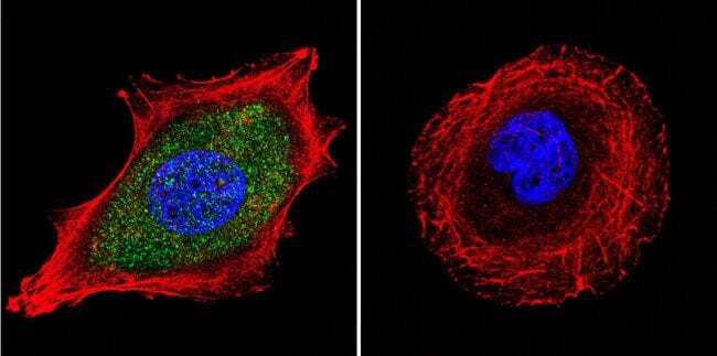
- Immunofluorescent analysis of Estrogen Receptor alpha using Anti-Estrogen Receptor alpha Monoclonal Antibody (EVG F9) (Product # MA3-310) shows staining in U251 Cells. Estrogen Receptor alpha staining (green), F-Actin staining with Phalloidin (red) and nuclei with DAPI (blue) is shown. Cells were grown on chamber slides and fixed with formaldehyde prior to staining. Cells were probed without (control) or with or an antibody recognizing Estrogen Receptor alpha (Product # MA3-310) at a dilution of 1:200 over night at 4°C, washed with PBS and incubated with a DyLight-488 conjugated secondary antibody (Product # 35503, Goat Anti-Mouse). Images were taken at 60X magnification.
- Submitted by
- Invitrogen Antibodies (provider)
- Main image

- Experimental details
- Immunofluorescent analysis of Estrogen Receptor alpha using Anti-Estrogen Receptor alpha Monoclonal Antibody (EVG F9) (Product # MA3-310) shows staining in MCF-7 Cells. Estrogen Receptor alpha staining (green), F-Actin staining with Phalloidin (red) and nuclei with DAPI (blue) is shown. Cells were grown on chamber slides and fixed with formaldehyde prior to staining. Cells were probed without (control) or with or an antibody recognizing Estrogen Receptor alpha (Product # MA3-310) at a dilution of 1:200 over night at 4°C, washed with PBS and incubated with a DyLight-488 conjugated secondary antibody (Product # 35503, Goat Anti-Mouse). Images were taken at 60X magnification.

Supportive validation
- Submitted by
- Invitrogen Antibodies (provider)
- Main image
- Experimental details
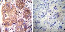
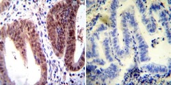
- Immunohistochemistry was performed on cancer biopsies of deparaffinized Human breast carcinoma tissues. To expose target proteins, heat induced antigen retrieval was performed using 10mM sodium citrate (pH6.0) buffer, microwaved for 8-15 minutes. Following antigen retrieval tissues were blocked in 3% BSA-PBS for 30 minutes at room temperature. Tissues were then probed at a dilution of 1:20 with a mouse monoclonal antibody recognizing Estrogen Receptor alpha (Product # MA3-310) or without primary antibody (negative control) overnight at 4°C in a humidified chamber. Tissues were washed extensively with PBST and endogenous peroxidase activity was quenched with a peroxidase suppressor. Detection was performed using a biotin-conjugated secondary antibody and SA-HRP, followed by colorimetric detection using DAB. Tissues were counterstained with hematoxylin and prepped for mounting.
- Submitted by
- Invitrogen Antibodies (provider)
- Main image
- Experimental details
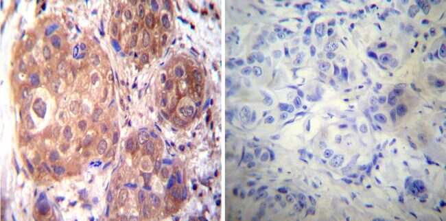
- Immunohistochemistry was performed on normal deparaffinized Human uterus tissue tissues. To expose target proteins, heat induced antigen retrieval was performed using 10mM sodium citrate (pH6.0) buffer, microwaved for 8-15 minutes. Following antigen retrieval tissues were blocked in 3% BSA-PBS for 30 minutes at room temperature. Tissues were then probed at a dilution of 1:100 with a mouse monoclonal antibody recognizing Estrogen Receptor alpha (Product # MA3-310) or without primary antibody (negative control) overnight at 4°C in a humidified chamber. Tissues were washed extensively with PBST and endogenous peroxidase activity was quenched with a peroxidase suppressor. Detection was performed using a biotin-conjugated secondary antibody and SA-HRP, followed by colorimetric detection using DAB. Tissues were counterstained with hematoxylin and prepped for mounting.

Supportive validation
- Submitted by
- Invitrogen Antibodies (provider)
- Main image
- Experimental details
- NULL

- Submitted by
- Invitrogen Antibodies (provider)
- Main image
- Experimental details
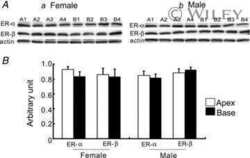
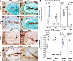
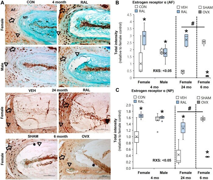
- FIGURE 1 Raloxifene increased ER-alpha protein expression in young and old IVDs, whereas OVX reduced ER-alpha protein expression. (A) Estrogen receptor-alpha (ER-alpha) immunostaining (arrow: presence of stain, arrowhead: absence of stain) in the (B) AF and (C) NP of young-adult (4 months), old (24 months), and OVX (6 months) mice. Data are represented as box plots with mean marked as cross (x), 25/75% deviation lines, and maximum/minimum whiskers. *: control (CON, n = 5/sex/group) vs. raloxifene (RAL); R: CON vs. RAL; S: male vs. female; RxS: interaction, vehicle (VEH, n = 8/group) vs. RAL, SHAM ( n = 4-5/group) vs. ovariectomized (OVX); # : aging effect (4 months vs. 6 months vs. 24 months), p < 0.05. AF, annulus fibrosus; NP, nucleus pulposus. Scale: 100 mum.
- Submitted by
- Invitrogen Antibodies (provider)
- Main image

- Experimental details
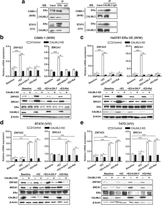
- Fig. 2 CALML3 as a coregulator of ERalpha affects the expression of ZNF423 and BRCA1. a Interaction of ERalpha and CALML3 in ZNF423 rs9940645 WT or variant genotype breast cancer cells treated with 0.01 nM E2. Co-IP performed by immunoprecipitating the protein complex with anti-ERalpha antibody, followed by immunoblotting for CALML3, or vice versa. b - e CALML3 was knocked down in ERalpha + breast cancer cells homozygous for ZNF423 WT or variant SNP genotypes and followed by treatment with 0.01 nM E2 +- 4-OH-TAM/raloxifene 10 -7 mol/L. ZNF423 and BRCA1mRNA expression levels relative to beta-ACTN shown as the mean of three independent experiments (+- SEM). * p < 0.05, ** p < 0.01, *** p < 0.001, **** p < 0.0001 by two-tailed Student's t test. Protein levels determined by western blot ( WB ) analysis. IP immunoprecipitation, 4-OH-TAM 4-hydroxytamoxifene, E2 estradiol, ER alpha estrogen receptor alpha, Ral raloxifene