Explore
 Validate
 Learn
Learn Western blot
Antibody data
- Antibody Data
- Antigen structure
- References [4]
- Comments [0]
- Validations
- Western blot [3]
- Immunocytochemistry [1]
- Immunohistochemistry [1]
- Other assay [3]
Submit
Validation data
Reference
Comment
Report error
- Product number
- PA5-16440 - Provider product page

- Provider
- Invitrogen Antibodies
- Product name
- Estrogen Receptor alpha Polyclonal Antibody
- Antibody type
- Polyclonal
- Antigen
- Synthetic peptide
- Description
- PA5-16440 targets Estrogen Receptor in IF, IHC (P), IP, and WB applications and shows reactivity with mouse, Hamster, and Human samples.
- Concentration
- 1 mg/mL
Submitted references Jie-Yu Pill, A Proprietary Herbal Medicine, Ameliorates Mood Disorder-Like Behavior and Cognitive Impairment in Estrogen-Deprived Mice Exposed to Chronic Unpredictable Mild Stress: Implication for a Potential Therapy of Menopause Syndrome.
Continuous Exposure of Breast Cancer Cells to Tamoxifen Upregulates GPER-1 and Increases Cell Proliferation.
27-hydroxycholesterol decreases cell proliferation in colon cancer cell lines.
ER-α36-mediated rapid estrogen signaling positively regulates ER-positive breast cancer stem/progenitor cells.
Zhou XD, Yang XJ, Zheng Y, Qin ZS, Sha W, Chen G, Zhang ZJ
Frontiers in psychiatry 2020;11:579995
Frontiers in psychiatry 2020;11:579995
Continuous Exposure of Breast Cancer Cells to Tamoxifen Upregulates GPER-1 and Increases Cell Proliferation.
Molina L, Bustamante F, Ortloff A, Ramos I, Ehrenfeld P, Figueroa CD
Frontiers in endocrinology 2020;11:563165
Frontiers in endocrinology 2020;11:563165
27-hydroxycholesterol decreases cell proliferation in colon cancer cell lines.
Warns J, Marwarha G, Freking N, Ghribi O
Biochimie 2018 Oct;153:171-180
Biochimie 2018 Oct;153:171-180
ER-α36-mediated rapid estrogen signaling positively regulates ER-positive breast cancer stem/progenitor cells.
Deng H, Zhang XT, Wang ML, Zheng HY, Liu LJ, Wang ZY
PloS one 2014;9(2):e88034
PloS one 2014;9(2):e88034
No comments: Submit comment
Supportive validation
- Submitted by
- Invitrogen Antibodies (provider)
- Main image

- Experimental details
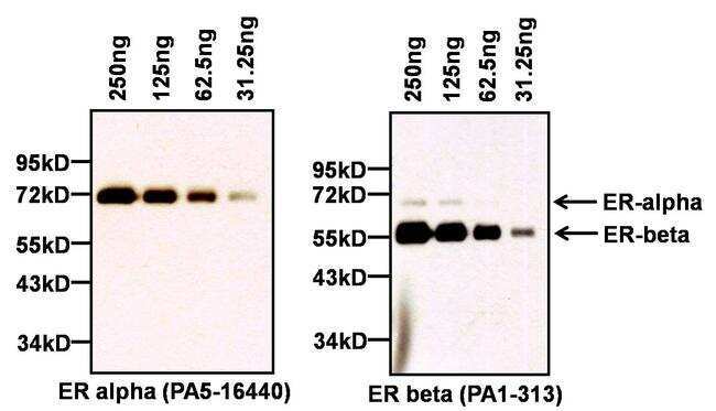
- Western blot analysis of Estrogen Receptor (ER) was performed by loading the indicated amounts of recombinant full-length human ER alpha (Product # A15674) and ER beta (Product # A15664) proteins, and 10 µL of PageRuler Prestained Protein Ladder (Product # 26616) per well onto a Novex® 4-20% Tris-Glycine polyacrylamide gel. Proteins were transferred to a PVDF membrane (Product # 88518) using the G2 Blotter (Product # 62288), and blocked with 5% milk in TBST for at least 1 hour at room temp. ER alpha was detected at ~66 kD (left panel) using an ER polyclonal antibody (Product # PA5-16440) at a dilution of 1:500 in blocking buffer overnight at 4C on a rocking platform, followed by an HRP-conjugated goat anti-rabbit IgG secondary antibody (Product # 31460) at a dilution of 1:40,000. Chemiluminescent detection was performed using SuperSignal West Pico (Product # 34080). The blot was then stripped with Restore Plus Western blot stripping buffer (Product # 46430) for at least 15 min at room temp, re-blocked with 5% milk in TBST for at least 1 hour at room temp, and re-probed with an ER beta polyclonal antibody (Product # PA1-313, right panel) at a dilution of 1:2000 overnight at 4C, followed by an HRP-conjugated goat anti-rabbit IgG secondary antibody (Product # 31460) at a dilution of 1:40,000 and chemiluminescent detection. The presence of faint ER-alpha bands on the blot probed with PA1-313 is due to incomplete stripping of PA5-16440.
- Submitted by
- Invitrogen Antibodies (provider)
- Main image
- Experimental details
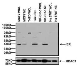
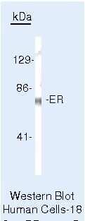
- Western blot analysis of Estrogen Receptor (ER) was performed by loading 20 µg of whole cell lysates (WCL) or nuclear extracts (NE) from the indicated breast cancer cell lines, and 10 µL of PageRuler Prestained Protein Ladder (Product # 26616) per well onto a Novex® 4-20% Tris-Glycine polyacrylamide gel. Proteins were transferred to a PVDF membrane (Product # 88518) using the G2 Blotter (Product # 62288), and blocked with 5% milk in TBST for at least 1 hour at room temp. ER was detected at ~43 kD (top panel) using an ER polyclonal antibody (Product # PA5-16440) at a dilution of 1:500 in blocking buffer overnight at 4C on a rocking platform, followed by an HRP-conjugated goat anti-rabbit IgG secondary antibody (Product # 31460) at a dilution of 1:40,000. Chemiluminescent detection was performed using SuperSignal West Dura (Product # 34075). To check for equivalent protein loading in each lane, the blot was stripped with Restore Plus Western blot stripping buffer (Product # 46430) for at least 15 min at room temp, re-blocked with 5% milk in TBST for at least 1 hour at room temp, and re-probed with an HDAC1 polyclonal antibody (Product # PA1-860, bottom panel) at a dilution of 1:1000 overnight at 4C, followed by an HRP-conjugated goat anti-rabbit IgG secondary antibody (Product # 31460) at a dilution of 1:40,000 and chemiluminescent detection.The molecular weight of the endogenous protein detected is consistent with isoforms of either ER alpha or ER beta.

- Submitted by
- Invitrogen Antibodies (provider)
- Main image
- Experimental details
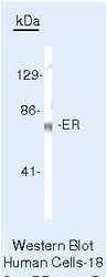
- Western blot of Estrogen Receptor using Estrogen Receptor Polyclonal Antibody (Product # PA5-16440) on MCF-7 Cells.
Supportive validation
- Submitted by
- Invitrogen Antibodies (provider)
- Main image

- Experimental details
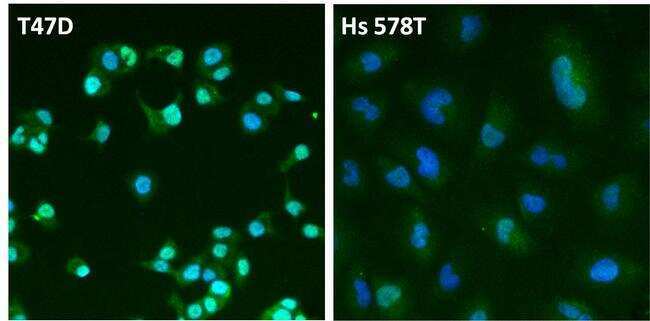
- Immunofluorescent analysis of Estrogen Receptor (ER, green) in T47D and Hs 578T breast cancer cells. The cells were fixed with 4% paraformaldehyde for 15 min at room temp, permeabilized with 0.1% Triton X-100 for 15 min at room temp, and blocked with 0.3% BSA in PBS for at least 30 min at room temp. Cells were stained with an ER polyclonal antibody (Product # PA5-16440) at a dilution of 1:100 in blocking buffer overnight at 4C, and then incubated with a DyLight 488-conjugated goat anti-rabbit IgG secondary antibody (Product # 35552) at a dilution of 1:500 for at least 1 hour at room temp. Nuclei (blue) were stained with DAPI (Product # 62247). Images were taken on a Thermo Scientific ToxInsight Instrument at 20X magnification.
Supportive validation
- Submitted by
- Invitrogen Antibodies (provider)
- Main image

- Experimental details
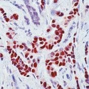
- Formalin-fixed, paraffin-embedded human breast carcinoma stained with ER, alpha antibody6 using peroxidase-conjugate and AEC chromogen. Note nuclear staining of tumor cells.
Supportive validation
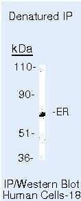
- Submitted by
- Invitrogen Antibodies (provider)
- Main image
- Experimental details
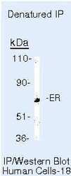
- Immunoprecipitation of Estrogen Receptor using Estrogen Receptor Polyclonal Antibody (Product # PA5-16440) on denatured Human MCF-7 Cells.
- Submitted by
- Invitrogen Antibodies (provider)
- Main image
- Experimental details
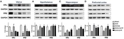
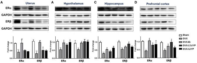
- Figure 10 Effects of 2.5 g/kg JYP (2.5 JYP), 5.0 g/kg JYP (5 JYP), and 0.3 mg/kg estradiol (E2) on the expression of ERalpha and ERbeta in the uterus (A) , hypothalamus (B) , hippocampus (C) , and prefrontal cortex (D) . Data are expressed as mean +- SEM ( n = 3) and examined with one-way analysis of variance (ANOVA), followed by post hoc between-group comparisons: * P < 0.05, ** P < 0.01, *** P < 0.001 vs. sham group; # P < 0.05, ## P < 0.01, vs. vehicle group.
- Submitted by
- Invitrogen Antibodies (provider)
- Main image

- Experimental details
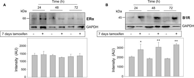
- Figure 5 Breast cancer cells continuously treated with tamoxifen overexpress the kinin B1R, but not ERalpha. MCF-7 cells that were under treatment with tamoxifen for 7 days together with their respective untreated controls were stimulated with 1,000 nM tamoxifen by 24, 48, and 72 h. Cell proteins were separated by SDS-PAGE, transferred onto Immobilon-P and immunoblotted with antibodies for detection of ERalpha (A) and kinin B1R (B) . Antibodies were stripped off and the same membranes were incubated with an antibody directed to GAPDH as control for protein loading. Data are representative of two independent experiments (n = 2). *P < 0.05; **P < 0.001 between tamoxifen-treated and untreated cells.