Explore
 Validate
 Learn
Learn Immunocytochemistry
Antibody data
- Antibody Data
- Antigen structure
- References [1]
- Comments [0]
- Validations
- Immunocytochemistry [1]
Submit
Validation data
Reference
Comment
Report error
- Product number
- MA5-13032 - Provider product page

- Provider
- Invitrogen Antibodies
- Product name
- ErbB2 (HER-2) Monoclonal Antibody (N28)
- Antibody type
- Monoclonal
- Antigen
- Other
- Description
- MA5-13032 targets HER-2 in IP applications and shows reactivity with Human samples.
- Antibody clone number
- N28
- Concentration
- 0.2 mg/mL
Submitted references Clathrin-independent endocytosis of ErbB2 in geldanamycin-treated human breast cancer cells.
Barr DJ, Ostermeyer-Fay AG, Matundan RA, Brown DA
Journal of cell science 2008 Oct 1;121(Pt 19):3155-66
Journal of cell science 2008 Oct 1;121(Pt 19):3155-66
No comments: Submit comment
Supportive validation
- Submitted by
- Invitrogen Antibodies (provider)
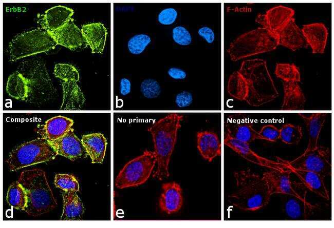
- Main image

- Experimental details
- Immunofluorescence analysis of Her2 was performed using 70% confluent log phase SK-BR-3 cells. The cells were fixed with 4% paraformaldehyde for 10 minutes, permeabilized with 0.1% Triton™ X-100 for 10 minutes, and blocked with 1% BSA for 1 hour at room temperature. The cells were labeled with Her2 Mouse monoclonal Antibody (Product # MA5-13032) at 5 µg/mL in 0.1% BSA and incubated overnight at 4 degree Celsius and then labeled with Goat anti-Mouse IgG (H+L) Superclonal™ Secondary Antibody, Alexa Fluor® 488 conjugate (Product # A28175) at a dilution of 1:2000 for 45 minutes at room temperature (Panel a: green). Nuclei (Panel b: blue) were stained with SlowFade® Gold Antifade Mountant with DAPI (Product # S36938). F-actin (Panel c: red) was stained with Rhodamine Phalloidin (Product # R415, 1:300). Panel d represents the merged image showing membranous and cytoplasmic localization. Panel f represents MDAMB-231 cells as negative controls, showing no Her2 staining. Panel e represents control cells with no primary antibody to assess background. The images were captured at 60X magnification.