 Explore
 Validate
 Learn
Learn Western blot
Antibody data
- Antibody Data
- Antigen structure
- References [1]
- Comments [0]
- Validations
- Western blot [2]
- Other assay [1]
Submit
Validation data
Reference
Comment
Report error
- Product number
- MA5-15050 - Provider product page

- Provider
- Invitrogen Antibodies
- Product name
- ErbB2 (HER-2) Monoclonal Antibody (K.929.9)
- Antibody type
- Monoclonal
- Antigen
- Synthetic peptide
- Description
- It is not recommended to aliquot this antibody.
- Antibody clone number
- K.929.9
- Concentration
- 256 µg/mL
Submitted references Constitutively activated PI3K accelerates tumor initiation and modifies histopathology of breast cancer.
Sheen MR, Marotti JD, Allegrezza MJ, Rutkowski M, Conejo-Garcia JR, Fiering S
Oncogenesis 2016 Oct 31;5(10):e267
Oncogenesis 2016 Oct 31;5(10):e267
No comments: Submit comment
Supportive validation
- Submitted by
- Invitrogen Antibodies (provider)
- Main image
- Experimental details
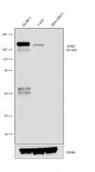
- Western blot analysis was performed on membrane enriched extracts (30 µg lysate) of SK-BR-3 (1), T-47D (2) and MDA-MB-231 (3). The blot was probed with anti-ErbB2 Rabbit Monoclonal Antibody (Product # MA5-15050, 1:1000 dilution) and detected by chemiluminescence using Goat anti-Rabbit IgG (H+L) Superclonal™ Secondary Antibody, HRP conjugate (Product # A27036, 0.25 µg/mL, 1:4000 dilution). A 185 kDa band corresponding to ErbB2 was observed in SK-BR-3 and moderate in T-47D where as it was not observed in MDA-MB-231 which is an ErbB2 negative cell line. Known quantity of protein samples were electrophoresed using Novex® NuPAGE® 4-12 % Bis-Tris gel (Product # NP0321BOX), XCell SureLock™ Electrophoresis System (Product # EI0002) and Novex® Sharp Pre-Stained Protein Standard (Product # LC5800). Resolved proteins were then transferred onto a nitrocellulose membrane with overnight wet transfer system. The membrane was probed with the relevant primary and secondary Antibody following blocking with 5 % skimmed milk. Chemiluminescent detection was performed using Pierce™ ECL Western Blotting Substrate (Product # 32106).

- Submitted by
- Invitrogen Antibodies (provider)
- Main image

- Experimental details
- Western blot analysis was performed on membrane enriched extracts (30 µg lysate) of SK-BR-3 (1), T-47D (2) and MDA-MB-231 (3). The blot was probed with anti-ErbB2 Rabbit Monoclonal Antibody (Product # MA5-15050, 1:1000 dilution) and detected by chemiluminescence using Goat anti-Rabbit IgG (H+L) Superclonal™ Secondary Antibody, HRP conjugate (Product # A27036, 0.25 µg/mL, 1:4000 dilution). A 185 kDa band corresponding to ErbB2 was observed in SK-BR-3 and moderate in T-47D where as it was not observed in MDA-MB-231 which is an ErbB2 negative cell line. Known quantity of protein samples were electrophoresed using Novex® NuPAGE® 4-12 % Bis-Tris gel (Product # NP0321BOX), XCell SureLock™ Electrophoresis System (Product # EI0002) and Novex® Sharp Pre-Stained Protein Standard (Product # LC5800). Resolved proteins were then transferred onto a nitrocellulose membrane with overnight wet transfer system. The membrane was probed with the relevant primary and secondary Antibody following blocking with 5 % skimmed milk. Chemiluminescent detection was performed using Pierce™ ECL Western Blotting Substrate (Product # 32106).

Supportive validation
- Submitted by
- Invitrogen Antibodies (provider)
- Main image
- Experimental details
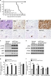
- Figure 1 In vivo expression of myr- p110alpha and homozygous p53 deletion in milk duct activates PI3K signaling and develops mammary tumors. ( a ) Kaplan-Meier survival curve demonstrating breast tumor onset defined as the first palpation-mediated recognition of tumors ( n =10 per group). Log-rank (Mantel-Cox) test was used for statistical analysis. *** P
