 Explore
 Validate
 Learn
Learn Western blot
Antibody data
- Antibody Data
- Antigen structure
- References [0]
- Comments [0]
- Validations
- Western blot [1]
- Immunocytochemistry [1]
Submit
Validation data
Reference
Comment
Report error
- Product number
- ABIN2508064 - Provider product page

- Provider
- antibodies-online
- Product name
- anti-V-Erb-B2 erythroblastic Leukemia Viral Oncogene Homolog 2, Neuro/glioblastoma Derived Oncogene Homolog (Avian) (ERBB2) (pTyr1222) antibody
- Antibody type
- Polyclonal
- Antigen
- This ERBB2 Antibody is generated from rabbits immunized with a KLH conjugated synthetic phosphopeptide corresponding to amino acid residues surrounding Y1222 of human ERBB2.Antigen type: Synthetic Peptide
- Description
- This antibody is purified through a protein A column, followed by peptide affinity purification.
- Reactivity
- Human
- Host
- Rabbit
- Epitope
- pTyr1222
- Vial size
- 200 μL
- Storage
- Maintain refrigerated at 2-8°C for up to 6 months. For long term storage store at -20°C in small aliquots to prevent freeze-thaw cycles.
No comments: Submit comment
Supportive validation
- Submitted by
- antibodies-online (provider)
- Main image
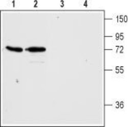
- Experimental details
- Western blot analysis of rat (lanes 1 and 3) and mouse brain membranes (lanes 2 and 4): 1, 2. Anti-H3 Histamine Receptor antibody (ABIN2510989), (1:200). 3, 4. Anti-H3 Histamine Receptor antibody, preincubated with the control peptide antigen. Expression of H3 Histamine Receptor in rat cerebellum Immunohistochemical staining of rat brain frozen sections with Anti-H3 Histamine Receptor antibody (ABIN2510989), (1:100), (green). A. H3 Histamine Receptor is particularly expressed in dendrites of Purkinje cells (arrows). B. Staining with mouse anti-parvalbumin (red) detected Purkinje cells and interneurons in the molecular layer. C. Merge of the two images demonstrates that the staining was restricted to dendrites of Purkinje cells. Cell nuclei were labeled with DAPI (blue) as the counterstain.

Supportive validation
- Submitted by
- antibodies-online (provider)
- Main image

- Experimental details
- Image(s): Immunofluorescence
