 Explore
 Validate
 Learn
Learn Western blot
 Immunoprecipitation
Antibody data
- Antibody Data
- Antigen structure
- References [3]
- Comments [0]
- Validations
- Immunoprecipitation [1]
- Immunohistochemistry [1]
- Other assay [1]
Submit
Validation data
Reference
Comment
Report error
- Product number
- PA5-16305 - Provider product page

- Provider
- Invitrogen Antibodies
- Product name
- ErbB2 (HER-2) Polyclonal Antibody
- Antibody type
- Polyclonal
- Antigen
- Synthetic peptide
- Description
- PA5-16305 targets HER-2 in IHC (P), IP, and WB applications and shows reactivity with mouse, Non-human primate, Rat, and Human samples. The PA5-16305 immunogen is synthetic peptide derived from C-terminus of human c-erbB-2/HER-2 protein. This Sequence is identical in rat neu protein.
- Reactivity
- Human, Mouse, Rat
- Host
- Rabbit
- Isotype
- IgG
- Vial size
- 500 μL
- Concentration
- 1 mg/mL
- Storage
- 4°C
Submitted references A Microfluidic Immunostaining System Enables Quality Assured and Standardized Immunohistochemical Biomarker Analysis.
The use of mouse models of breast cancer and quantitative image analysis to evaluate hormone receptor antigenicity after microwave-assisted formalin fixation.
The mucin MUC4 and its membrane partner ErbB2 regulate biological properties of human CAPAN-2 pancreatic cancer cells via different signalling pathways.
Kwon S, Cho CH, Kwon Y, Lee ES, Park JK
Scientific reports 2017 Apr 5;7:45968
Scientific reports 2017 Apr 5;7:45968
The use of mouse models of breast cancer and quantitative image analysis to evaluate hormone receptor antigenicity after microwave-assisted formalin fixation.
Engelberg JA, Giberson RT, Young LJ, Hubbard NE, Cardiff RD
The journal of histochemistry and cytochemistry : official journal of the Histochemistry Society 2014 May;62(5):319-34
The journal of histochemistry and cytochemistry : official journal of the Histochemistry Society 2014 May;62(5):319-34
The mucin MUC4 and its membrane partner ErbB2 regulate biological properties of human CAPAN-2 pancreatic cancer cells via different signalling pathways.
Jonckheere N, Skrypek N, Merlin J, Dessein AF, Dumont P, Leteurtre E, Harris A, Desseyn JL, Susini C, Frénois F, Van Seuningen I
PloS one 2012;7(2):e32232
PloS one 2012;7(2):e32232
No comments: Submit comment
Supportive validation
- Submitted by
- Invitrogen Antibodies (provider)
- Main image

- Experimental details
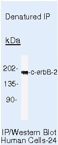
- Immunoprecipitation of HER-2 using HER-2 Polyclonal Antibody (Product # PA5-16305) on denatured Human SKBR3 Cells.
Supportive validation
- Submitted by
- Invitrogen Antibodies (provider)
- Main image
- Experimental details
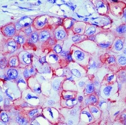
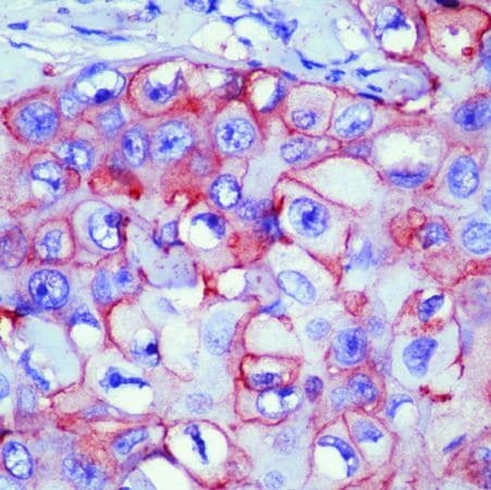
- Formalin-fixed, paraffin-embedded human breast cancer stained with c-erbB2 antibody using peroxidase-conjugate and AEC chromogen. Note membrane staining of tumor cells.
Supportive validation
- Submitted by
- Invitrogen Antibodies (provider)
- Main image
- Experimental details
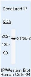
- Immunoprecipitation of HER-2 using HER-2 Polyclonal Antibody (Product # PA5-16305) on denatured Human SKBR3 Cells.
