Explore
 Validate
 Learn
Learn Western blot
Antibody data
- Antibody Data
- Antigen structure
- References [1]
- Comments [0]
- Validations
- Western blot [2]
- Immunocytochemistry [1]
- Immunohistochemistry [1]
Submit
Validation data
Reference
Comment
Report error
- Product number
- PA5-16774 - Provider product page

- Provider
- Invitrogen Antibodies
- Product name
- ErbB2 (HER-2) Polyclonal Antibody
- Antibody type
- Polyclonal
- Antigen
- Synthetic peptide
- Description
- PA5-16774 targets HER-2 in IP and WB applications and shows reactivity with Human, mouse, Non-human primate, and Rat samples. The PA5-16774 immunogen is a synthetic peptide from C-terminus of human HER-2 protein. This sequence is identical in rat neu protein.
- Reactivity
- Human, Mouse, Rat
- Host
- Rabbit
- Isotype
- IgG
- Vial size
- 500 µL
- Storage
- -20° C, Avoid Freeze/Thaw Cycles
Submitted references erbB3 is an active tyrosine kinase capable of homo- and heterointeractions.
Steinkamp MP, Low-Nam ST, Yang S, Lidke KA, Lidke DS, Wilson BS
Molecular and cellular biology 2014 Mar;34(6):965-77
Molecular and cellular biology 2014 Mar;34(6):965-77
No comments: Submit comment
Supportive validation
- Submitted by
- Invitrogen Antibodies (provider)
- Main image

- Experimental details
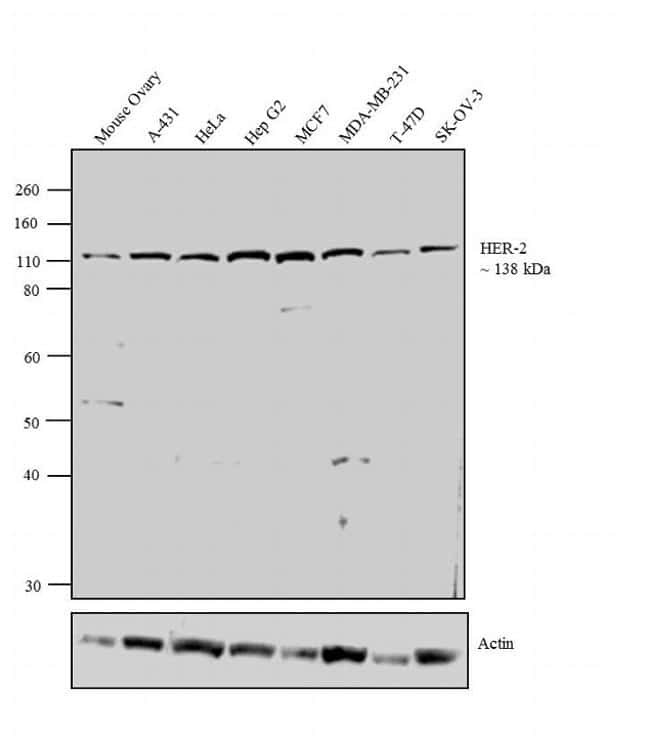
- Western blot analysis was performed on tissue extract (30 µg lysate) of Mouse Ovary (Lane 1), whole cell extracts (30 µg lysate) of A-431 (Lane 2), HeLa (Lane 3), Hep G2 (Lane 4), MCF7 (Lane 5), MDA-MB-231 (Lane 6), T-47D (Lane 7) and SK-OV-3 (Lane 8). The blots were probed with Anti-HER-2 Rabbit Polyclonal Antibody (Product # PA5-16774, 1-3 µg/mL) and detected by chemiluminescence using Goat anti-Rabbit IgG (H+L) Superclonal™ Secondary Antibody, HRP conj µgate (Product # A27036, 0.4 µg/mL, 1:2500 dilution). A 138 kDa band corresponding to HER-2 was observed across the cell lines tested. Known quantity of protein samples were electrophoresed using Novex® NuPAGE® 10 % Bis-Tris gel (Product # NP0302BOX), XCell SureLock™ Electrophoresis System (Product # EI0002) and Novex® Sharp Pre-Stained Protein Standard (Product # LC5800). Resolved proteins were then transferred onto a nitrocellulose membrane with overnight wet transfer system. The membrane was probed with the relevant primary and secondary Antibody following blocking with 5 % skimmed milk. Chemiluminescent detection was performed using Pierce™ ECL Western Blotting Substrate (Product # 32106).

- Submitted by
- Invitrogen Antibodies (provider)
- Main image

- Experimental details
- Western blot analysis was performed on tissue extract (30 µg lysate) of Mouse Ovary (Lane 1), whole cell extracts (30 µg lysate) of A-431 (Lane 2), HeLa (Lane 3), Hep G2 (Lane 4), MCF7 (Lane 5), MDA-MB-231 (Lane 6), T-47D (Lane 7) and SK-OV-3 (Lane 8). The blots were probed with Anti-HER-2 Rabbit Polyclonal Antibody (Product # PA5-16774, 1-3 µg/mL) and detected by chemiluminescence using Goat anti-Rabbit IgG (H+L) Superclonal™ Secondary Antibody, HRP conj µgate (Product # A27036, 0.4 µg/mL, 1:2500 dilution). A 138 kDa band corresponding to HER-2 was observed across the cell lines tested. Known quantity of protein samples were electrophoresed using Novex® NuPAGE® 10 % Bis-Tris gel (Product # NP0302BOX), XCell SureLock™ Electrophoresis System (Product # EI0002) and Novex® Sharp Pre-Stained Protein Standard (Product # LC5800). Resolved proteins were then transferred onto a nitrocellulose membrane with overnight wet transfer system. The membrane was probed with the relevant primary and secondary Antibody following blocking with 5 % skimmed milk. Chemiluminescent detection was performed using Pierce™ ECL Western Blotting Substrate (Product # 32106).
Supportive validation
- Submitted by
- Invitrogen Antibodies (provider)
- Main image
- Experimental details
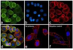
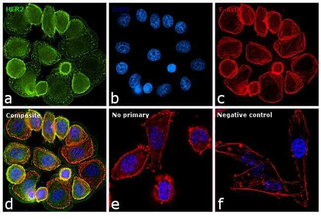
- Immunofluorescence analysis of Her2 was performed using 70% confluent log phase SK-BR-3 cells. The cells were fixed with 4% paraformaldehyde for 10 minutes, permeabilized with 0.1% Triton™ X-100 for 10 minutes, and blocked with 1% BSA for 1 hour at room temperature. The cells were labeled with Her2 Rabbit polyclonal Antibody (Product # PA5-16774) at 5 µg/mL in 0.1% BSA and incubated overnight at 4 degree Celsius and then labeled with Goat anti-Rabbit IgG (H+L) Superclonal™ Secondary Antibody, Alexa Fluor® 488 conjugate (Product # A27034) at a dilution of 1:2000 for 45 minutes at room temperature (Panel a: green). Nuclei (Panel b: blue) were stained with SlowFade® Gold Antifade Mountant with DAPI (Product # S36938). F-actin (Panel c: red) was stained with Rhodamine Phalloidin (Product # R415, 1:300). Panel d represents the merged image showing membranous and cytoplasmic localization. Panel f represents MDAMB-231 cells as negative controls, showing no Her2 staining. Panel e represents control cells with no primary antibody to assess background. The images were captured at 60X magnification.
Supportive validation
- Submitted by
- Invitrogen Antibodies (provider)
- Main image
- Experimental details
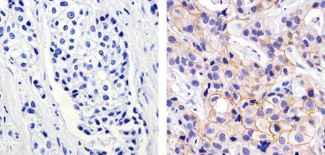
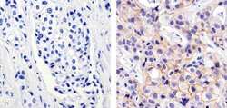
- Immunohistochemistry analysis of HER-2/ErbB2 showing staining in the membrane of paraffin-embedded human breast carcinoma (right) compared to a negative control without primary antibody (left). To expose target proteins, antigen retrieval was performed using 10mM sodium citrate (pH 6.0), microwaved for 8-15 min. Following antigen retrieval, tissues were blocked in 3% H2O2-methanol for 15 min at room temperature, washed with ddH2O and PBS, and then probed with a HER-2/ErbB2 Rabbit Polyclonal Antibody (Product # PA5-16774) diluted in 3% BSA-PBS at a dilution of 1:100 for 1 hour at 37°C in a humidified chamber. Tissues were washed extensively in PBST and detection was performed using an HRP-conjugated secondary antibody followed by colorimetric detection using a DAB kit. Tissues were counterstained with hematoxylin and dehydrated with ethanol and xylene to prep for mounting.