Explore
 Validate
 Learn
Learn Western blot
 Immunocytochemistry
Antibody data
- Antibody Data
- Antigen structure
- References [3]
- Comments [0]
- Validations
- Immunocytochemistry [1]
- Immunohistochemistry [1]
Submit
Validation data
Reference
Comment
Report error
- Product number
- BAF1129 - Provider product page

- Provider
- R&D Systems
- Product name
- Human ErbB2/Her2 Biotinylated Antibody
- Antibody type
- Polyclonal
- Description
- Antigen Affinity-purified. Detects human ErbB2 in ELISAs and Western blots. In sandwich immunoassays, less than 0.3% cross-reactivity with recombinant human (rh) ErbB3, rhErbB4, and rhEGF R is observed.
- Reactivity
- Human
- Host
- Goat
- Conjugate
- Biotin
- Antigen sequence
P04626- Isotype
- IgG
- Vial size
- 50 ug
- Concentration
- LYOPH
- Storage
- Use a manual defrost freezer and avoid repeated freeze-thaw cycles. 12 months from date of receipt, -20 to -70 °C as supplied. 1 month, 2 to 8 °C under sterile conditions after reconstitution. 6 months, -20 to -70 °C under sterile conditions after reconstitution.
Submitted references Molecular nanoshearing: an innovative approach to shear off molecules with AC-induced nanoscopic fluid flow.
Microbead arrays for the analysis of ErbB receptor tyrosine kinase activation and dimerization in breast cancer cells.
Development and validation of sandwich ELISA microarrays with minimal assay interference.
Shiddiky MJ, Vaidyanathan R, Rauf S, Tay Z, Trau M
Scientific reports 2014 Jan 16;4:3716
Scientific reports 2014 Jan 16;4:3716
Microbead arrays for the analysis of ErbB receptor tyrosine kinase activation and dimerization in breast cancer cells.
Khan IH, Zhao J, Ghosh P, Ziman M, Sweeney C, Kung HJ, Luciw PA
Assay and drug development technologies 2010 Feb;8(1):27-36
Assay and drug development technologies 2010 Feb;8(1):27-36
Development and validation of sandwich ELISA microarrays with minimal assay interference.
Gonzalez RM, Seurynck-Servoss SL, Crowley SA, Brown M, Omenn GS, Hayes DF, Zangar RC
Journal of proteome research 2008 Jun;7(6):2406-14
Journal of proteome research 2008 Jun;7(6):2406-14
No comments: Submit comment
Supportive validation
- Submitted by
- R&D Systems (provider)
- Main image

- Experimental details
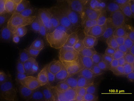
- ErbB2/Her2 in MCF-7 Human Cell Line. ErbB2/Her2 was detected in immersion fixed MCF-7 human breast cancer cell line using Goat Anti-Human ErbB2/Her2 Biotinylated Antigen Affinity-purified Polyclonal Antibody (Catalog # BAF1129) at 10 µg/mL for 3 hours at room temperature. Cells were stained using the NorthernLights™ 557-conjugated Streptavidin (yellow; Catalog # NL999) and counterstained with DAPI (blue). View our protocol for Fluorescent ICC Staining of Cells on Coverslips.
Supportive validation
- Submitted by
- R&D Systems (provider)
- Main image
- Experimental details
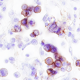
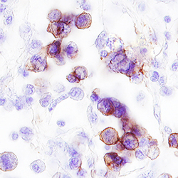
- ErbB2/Her2 in Human Breast Cancer Tissue. ErbB2/Her2 was detected in immersion fixed paraffin-embedded sections of human breast cancer tissue using Goat Anti-Human ErbB2/Her2 Biotinylated Antigen Affinity-purified Polyclonal Antibody (Catalog # BAF1129) at 15 µg/mL overnight at 4 °C. Tissue was stained using the Anti-Goat HRP-DAB Cell & Tissue Staining Kit (brown; Catalog # CTS008) and counterstained with hematoxylin (blue). Specific staining was localized to plasma membrane. View our protocol for Chromogenic IHC Staining of Paraffin-embedded Tissue Sections.