Explore
 Validate
 Learn
Learn Western blot
Antibody data
- Antibody Data
- Antigen structure
- References [0]
- Comments [0]
- Validations
- Western blot [1]
- Immunocytochemistry [1]
- Flow cytometry [1]
Submit
Validation data
Reference
Comment
Report error
- Product number
- 701640 - Provider product page

- Provider
- Invitrogen Antibodies
- Product name
- PSA Recombinant Rabbit Monoclonal Antibody (4H23L24)
- Antibody type
- Monoclonal
- Antigen
- Synthetic peptide
- Reactivity
- Human
- Host
- Rabbit
- Isotype
- IgG
- Antibody clone number
- 4H23L24
- Vial size
- 100 µg
- Concentration
- 0.5 mg/mL
- Storage
- Store at 4°C short term. For long term storage, store at -20°C, avoiding freeze/thaw cycles.
No comments: Submit comment
Supportive validation
- Submitted by
- Invitrogen Antibodies (provider)
- Main image
- Experimental details
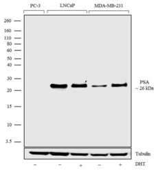
- Western blot analysis was performed on whole cell extracts (30 µg lysate) of PC3 (Lane 1), LnCAP (Lane 2), LnCAP treated with DHT (100nM DHT for 24hrs) (Lane 3), MDA-MB-231 (Lane 4) and MDA-MB-231 treated with DHT (100nM DHT for 24hrs) (Lane 5). The blots were probed with Anti-PSA Recombinant Rabbit Monoclonal Antibody (Product # 701640, 1-2 µg/mL) and detected by chemiluminescence using Goat anti-Rabbit IgG (H+L) Superclonal™ Secondary Antibody, HRP conjugate (Product # A27036, 0.4 µg/mL, 1:2500 dilution). A 26 kDa band corresponding to PSA was observed in LnCAP and and no band was observed in PC-3, as reported in literature. Known quantity of protein samples were electrophoresed using Novex® NuPAGE® 12% Bis-Tris gel (Product # NP0342BOX), XCell SureLock™ Electrophoresis System (Product # EI0002) and Novex® Sharp Pre-Stained Protein Standard (Product # LC5800). Resolved proteins were then transferred onto a nitrocellulose membrane using iBlot® Dry Blotting System (Product # IB21001). The membrane was probed with the relevant primary and secondary Antibody following blocking with 5% skimmed milk. Chemiluminescent detection was performed using Pierce™ ECL Western blotting Substrate (Product # 32106).
Supportive validation
- Submitted by
- Invitrogen Antibodies (provider)
- Main image

- Experimental details
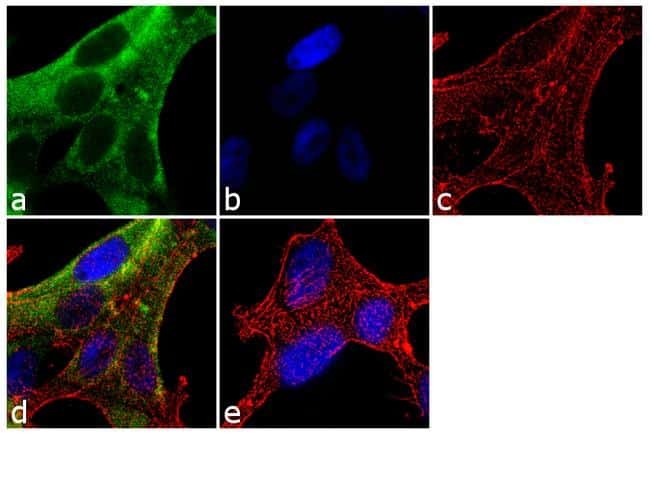
- Immunofluorescence was performed on fixed and permeabilized LNCaP cells for detection of PSA using PSA Recombinant Rabbit Monoclonal Antibody (Product # 701640, 2 µg/mL) and labeled with Goat anti-Rabbit IgG (H+L) Superclonal™ Secondary Antibody, Alexa Fluor® 488 conjugate (Product # A27034, 1:2000). Panel a) shows representative cells that were stained for detection and localization of PSA protein (green), Panel b) is stained for nuclei (blue) using SlowFade® Gold Antifade Mountant with DAPI (Product # S36938). Panel c) represents cytoskeletal F-actin staining using Alexa Fluor® 555 Rhodamine Phalloidin (Product # R415, 1:300). Panel d) is a composite image of Panels a, b and c clearly demonstrating cytoplasmic localization of PSA. Panel e) represents control cells with no primary antibody to assess background.
Supportive validation
- Submitted by
- Invitrogen Antibodies (provider)
- Main image
- Experimental details
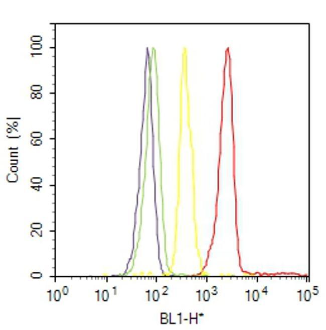
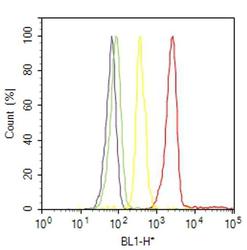
- Flow Cytometry analysis of PSA was performed on LNCaP cells treated with DHT (200nm for 24 hours) followed by GolgiStop™ treatment for 1 hour. Cells were labeled with ABfinity™ Anti-PSA Recombinant Rabbit Monoclonal Antibody (Product# 701640, 5 ug/ 1M cells) or with rabbit isotype control at 0.5 ug/ml and detected with Goat anti-Rabbit IgG (H+L) Superclonal™ Secondary Antibody, (Alexa Fluor® 488 conjugate, Product # A27034, 0.4 ug/ml, 1:2500) as represented by the red and yellow histograms respectively. The purple histogram represents unstained control cells and the green histogram represents no-primary-antibody control. A representative of 10,000 cells were acquired and analyzed for each sample using an Attune® Acoustic Focusing Cytometer (4468770).