Explore
 Validate
 Learn
Learn Western blot
Antibody data
- Antibody Data
- Antigen structure
- References [0]
- Comments [0]
- Validations
- Western blot [1]
- Immunocytochemistry [1]
- Immunohistochemistry [2]
Submit
Validation data
Reference
Comment
Report error
- Product number
- MA5-14470 - Provider product page

- Provider
- Invitrogen Antibodies
- Product name
- PSA Monoclonal Antibody (EP1588Y)
- Antibody type
- Monoclonal
- Antigen
- Synthetic peptide
- Description
- MA5-14470 targets Prostate Specific Antigen in IHC (P), ICC/IF and WB applications and shows reactivity with Human samples, however this antibody is not recommended for human colon and mouse prostate tissue sections. This antibody does not react with rat tissue in Western blot applications.
- Antibody clone number
- EP1588Y
- Concentration
- Conc. Not Determined
No comments: Submit comment
Supportive validation
- Submitted by
- Invitrogen Antibodies (provider)
- Main image

- Experimental details
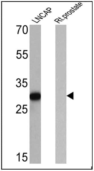
- Western blot analysis of Prostate Specific Antigen was performed by loading 25 µg of LNCAP (lane 1) and rat prostate (lane 2) cell lysates onto an SDS polyacrylamide gel. Proteins were transferred to a PVDF membrane and blocked at 4ºC overnight. The membrane was probed with a Prostate Specific Antigen monoclonal antibody (Product # MA5-14470) at a dilution of 1:50 overnight at 4°C, washed in TBST, and probed with an HRP-conjugated secondary antibody for 1 hr at room temperature in the dark. Chemiluminescent detection was performed using Pierce ECL Plus Western Blotting Substrate (Product # 32132). Results show a band at ~29 kDa in LNCAP cells.
Supportive validation
- Submitted by
- Invitrogen Antibodies (provider)
- Main image
- Experimental details
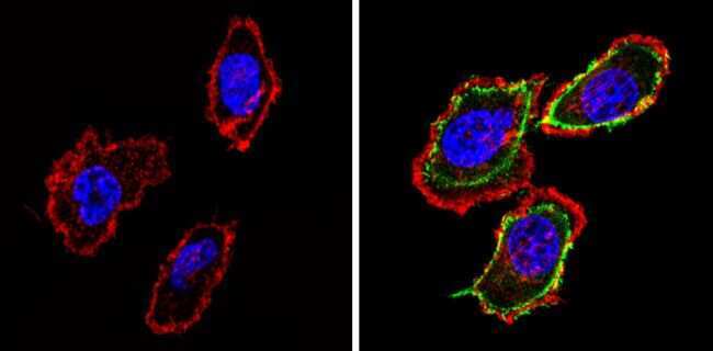
- Immunofluorescent analysis of Prostate Specific Antigen (green) showing staining in the cytoplasm of PC-3 cells (right) compared to a negative control without primary antibody (left). Formalin-fixed cells were permeabilized with 0.1% Triton X-100 in TBS for 5-10 minutes and blocked with 3% BSA-PBS for 30 minutes at room temperature. Cells were probed with a Prostate Specific Antigen monoclonal antibody (Product # MA5-14470) in 3% BSA-PBS at a dilution of 1:100 and incubated overnight at 4 ºC in a humidified chamber. Cells were washed with PBST and incubated with a DyLight-conjugated secondary antibody in PBS at room temperature in the dark. F-actin (red) was stained with a fluorescent red phalloidin and nuclei (blue) were stained with Hoechst or DAPI. Images were taken at a magnification of 60x.
Supportive validation
- Submitted by
- Invitrogen Antibodies (provider)
- Main image

- Experimental details
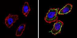
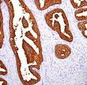
- Formalin-fixed, paraffin-embedded human prostate stained with rabbit monoclonal PSA using peroxidase-conjugate and DAB chromogen. Note cytoplasmic staining.
- Submitted by
- Invitrogen Antibodies (provider)
- Main image

- Experimental details
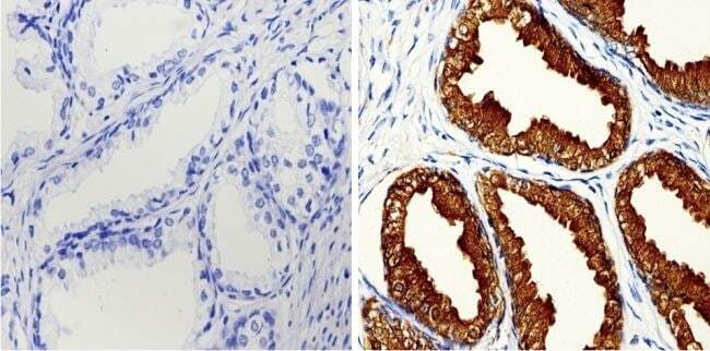
- Immunohistochemistry analysis of Prostate Specific Antigen showing positive staining in the Secretion of paraffin-treated Human prostate tissue (right) compared with a negative control in the absence of primary antibody (left). To expose target proteins, antigen retrieval method was performed using 10mM sodium citrate (pH 6.0) microwaved for 8-15 min. Following antigen retrieval, tissues were blocked in 3% H2O2-methanol for 15 min at room temperature, washed with ddH2O and PBS, and then probed with a Prostate Specific Antigen antibody (Product # MA5-14470) diluted by 3% BSA-PBS at a dilution of 1:20-1:100 overnight at 4°C in a humidified chamber. Tissues were washed extensively PBST and detection was performed using an HRP-conjugated secondary antibody followed by colorimetric detection using a DAB kit. Tissues were counterstained with hematoxylin and dehydrated with ethanol and xylene to prep for mounting.