Explore
 Validate
 Learn
LearnM01262
antibody from Boster Biological Technology
Targeting: PMEL
D12S53E, gp100, Pmel17, SI, SIL, SILV
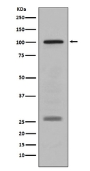
 Western blot
 Immunohistochemistry
Antibody data
- Antibody Data
- Antigen structure
- References [0]
- Comments [0]
- Validations
- Immunohistochemistry [1]
Submit
Validation data
Reference
Comment
Report error
- Product number
- M01262 - Provider product page

- Provider
- Boster Biological Technology
- Product name
- Anti-PMEL17 / GP100 Rabbit Monoclonal Antibody
- Antibody type
- Monoclonal
- Description
- Monoclonal antibody for PMEL17/PMEL detection. Host: Rabbit.Size: 100ug/vial. Tested applications: IHC, WB. Reactive species: Human PMEL17/PMEL information: Molecular Weight: 70255 MW; Subcellular Localization: Endoplasmic reticulum membrane; Single-pass type I membrane protein. Golgi apparatus. Melanosome. Endosome, multivesicular body. Identified by mass spectrometry in melanosome fractions from stage I to stage IV. Localizes predominantly to intralumenal vesicles (ILVs) within multivesicular bodies. Associates with ILVs found within the lumen of premelanosomes and melanosomes and particularly in compartments that serve as precursors to the striated stage II premelanosomes; Tissue Specificity: Preferentially expressed in melanomas. Some expression was found in dysplastic nevi. Not found in normal tissues nor in carcinomas. Normally expressed at low levels in quiescent adult melanocytes but overexpressed by proliferating neonatal melanocytes and during tumor growth.
- Reactivity
- Human
- Host
- Rabbit
- Antibody clone number
- AHH-16
- Vial size
- 100ug/vial
- Concentration
- 0.5-1mg/ml, actual concentration vary by lot. Use suggested dilution ratio to decide dilution procedure.
- Storage
- At -20°C for one year. Avoid repeated freezing and thawing.
No comments: Submit comment
Supportive validation
- Submitted by
- Boster Biological Technology (provider)
- Main image
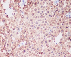
- Experimental details
- Immunohistochemical analysis of paraffin-embedded human melanoma, using PMEL17 / GP100 Antibody(M01262)PMEL was detected in paraffin-embedded tissue section. Heat mediated antigen retrieval was performed in citrate buffer (pH6, epitope retrieval solution) for 20 mins. The tissue section was blocked with 10% goat serum. The tissue section was then incubated with 1ug/ml rabbit anti-PMEL Antibody (M01262)overnight at 4?? Biotinylated goat anti-rabbit IgG was used as secondary antibody and incubated for 30 minutes at 37?? The tissue section was developed using Strepavidin-Biotin-Complex (SABC)(Catalog # SA1022) with DAB as the chromogen.
- Additional image