Explore
 Validate
 Learn
Learn Western blot
Antibody data
- Antibody Data
- Antigen structure
- References [1]
- Comments [0]
- Validations
- Western blot [2]
- Immunohistochemistry [1]
- Other assay [2]
Submit
Validation data
Reference
Comment
Report error
- Product number
- PA5-32491 - Provider product page

- Provider
- Invitrogen Antibodies
- Product name
- PMEL Polyclonal Antibody
- Antibody type
- Polyclonal
- Antigen
- Recombinant full-length protein
- Description
- Heat-mediated antigen retrieval is recommended prior to staining, using a 10mM citrate buffer, pH 6.0, for 10 minutes followed by cooling at room temperature for 20 min. Following antigen retrieval, incubate samples with primary antibody for 10 min at room temperature. A suggested positive control is melanoma.
- Reactivity
- Human
- Host
- Rabbit
- Isotype
- IgG
- Vial size
- 500 µL
- Concentration
- 0.123 mg/mL
- Storage
- Store at 4°C short term. For long term storage, store at -20°C, avoiding freeze/thaw cycles.
Submitted references Tissue-engineered 3D melanoma model with blood and lymphatic capillaries for drug development.
Bourland J, Fradette J, Auger FA
Scientific reports 2018 Sep 4;8(1):13191
Scientific reports 2018 Sep 4;8(1):13191
No comments: Submit comment
Supportive validation
- Submitted by
- Invitrogen Antibodies (provider)
- Main image

- Experimental details
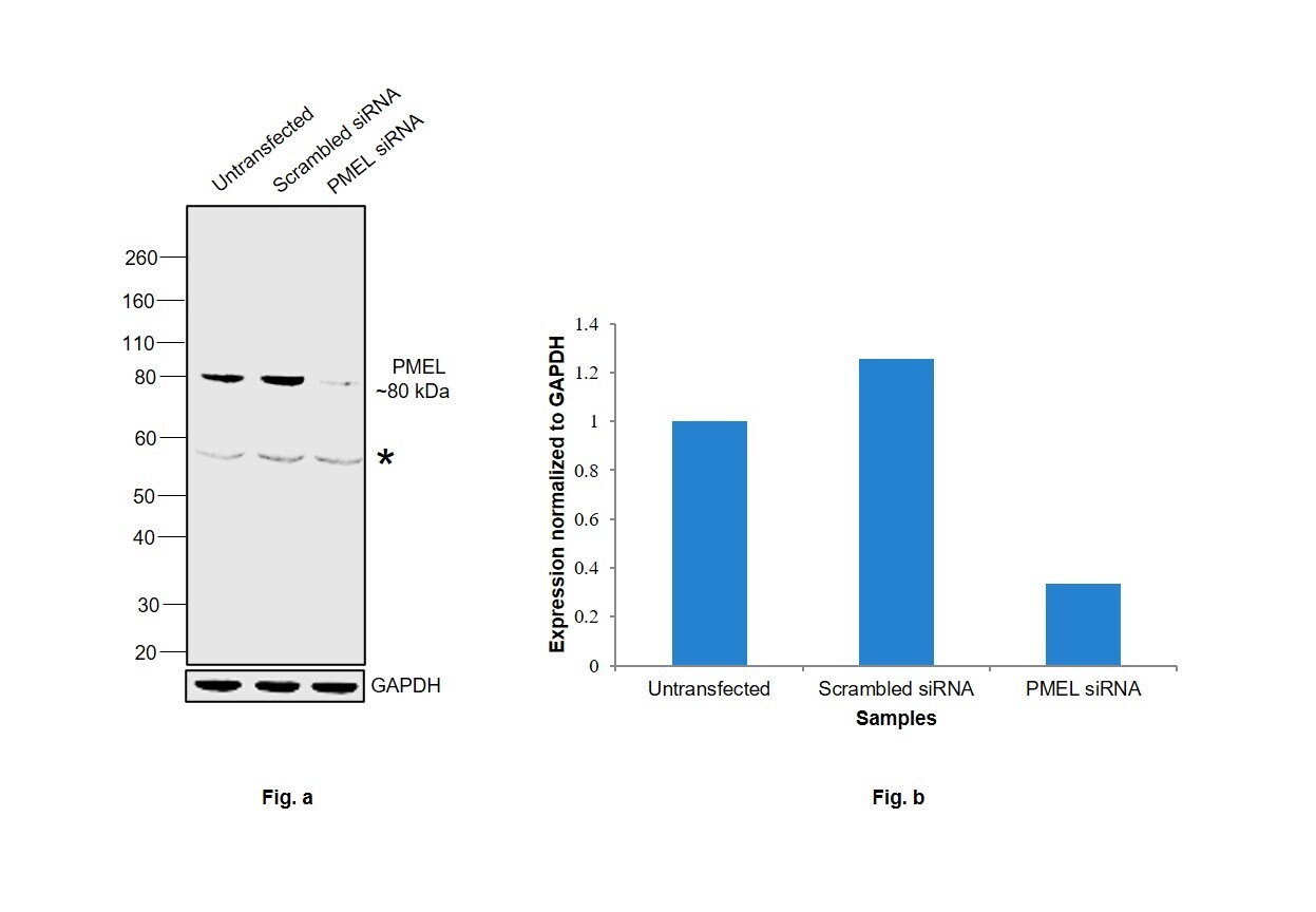
- Knockdown of PMEL was achieved by transfecting SK-MEL-5 with PMEL specific siRNAs (Silencer® select Product # s12861, s12859). Western blot analysis (Fig. a) was performed using Whole cell extracts from the PMEL knockdown cells (lane 3), non-targeting scrambled siRNA transfected cells (lane 2) and untransfected cells (lane 1). The blot was probed with PMEL Polyclonal Antibody (Product # PA5-32491, 1 µg/mL ) and Goat anti-Rabbit IgG (H+L) Superclonal™ Recombinant Secondary Antibody, HRP (Product # A27036, 1:4000 dilution). Densitometric analysis of this western blot is shown in histogram (Fig. b). The decrease in signal upon siRNA mediated knockdown confirms that antibody is specific to PMEL.
- Submitted by
- Invitrogen Antibodies (provider)
- Main image
- Experimental details
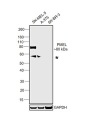
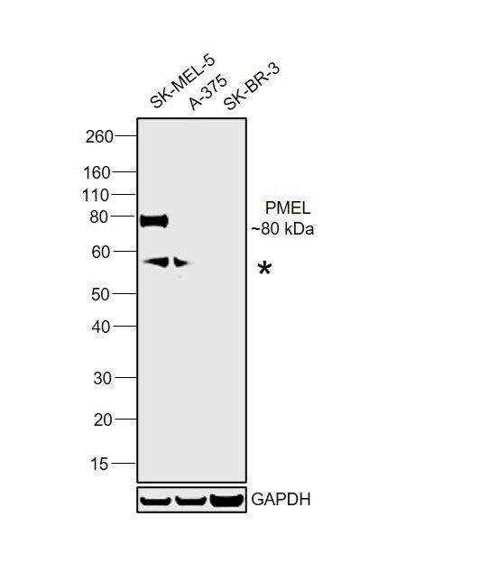
- Western blot was performed using Anti-PMEL Polyclonal Antibody (Product # PA5-32491) and an 80 kDa band corresponding to PMEL was observed across in SK-MEL-5 in comparison to A-375 and SK-BR-3 which is supposed to be negative. A non-specific band around 58 kDa was also observed. Whole cell extracts (30 µg lysate) of SK-MEL-5 (Lane 1), A-375 (Lane 2) and SK-BR-3 (Lane 3) were electrophoresed using NuPAGE™ 4-12% Bis-Tris Protein Gel (Product # NP0322BOX). Resolved proteins were then transferred onto a Nitrocellulose membrane (Product # IB23001) by iBlot® 2 Dry Blotting System (Product # IB21001). The blot was probed with the primary antibody (1 µg/mL) and detected by chemiluminescence with Goat anti-Rabbit IgG (H+L) Superclonal™ Recombinant Secondary Antibody, HRP (Product # A27036, 1:4000 dilution) using the iBright FL 1000 (Product # A32752). Chemiluminescent detection was performed using Novex® ECL Chemiluminescent Substrate Reagent Kit (Product # WP20005).
Supportive validation
- Submitted by
- Invitrogen Antibodies (provider)
- Main image
- Experimental details
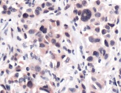
- Immunohistochemical analysis of Melanoma using anti-Melanoma Polyclonal Antibody (Product # PA5-32491) in Melanoma Cancer Tissue. The recommened dilution for this antibody in immunohistochemistry applications is 1:50.

Supportive validation
- Submitted by
- Invitrogen Antibodies (provider)
- Main image
- Experimental details
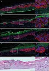
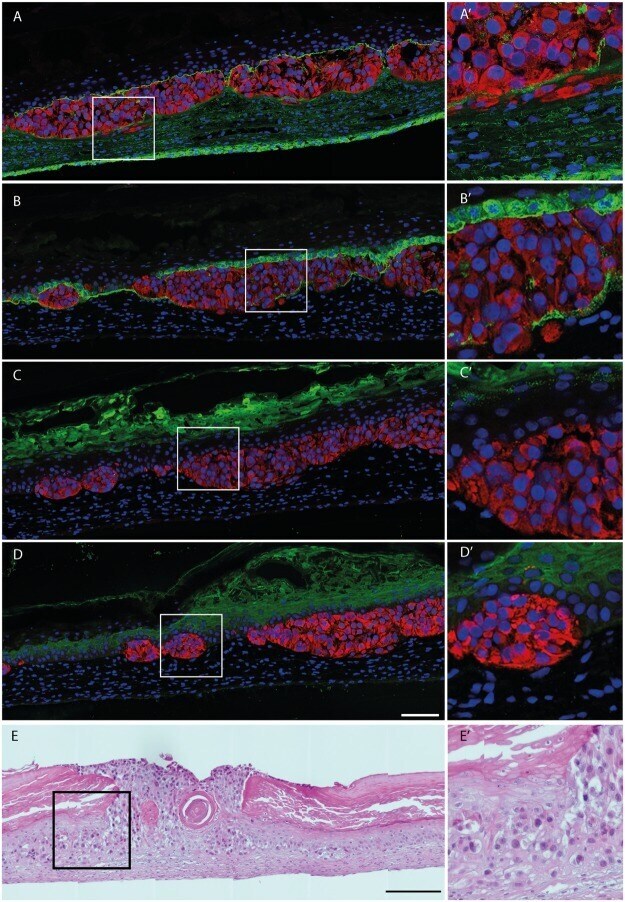
- Figure 5 Melanoma spheroids integration into the human reconstructed cutaneous microenvironment. ( A - D ) Immunostainings of selected cutaneous proteins (green signal): ( A ) fibronectin staining of the dermis, ( B ) laminin 332 staining indicating the presence of a basement membrane, ( C ) filaggrin and ( D ) involucrin stainings (scale bar: 100 um). Melanoma cells (SK-MEL 28) are labeled in red following premelanosome protein (PMEL) staining while nuclei are stained with Hoechst (blue signal). (A'-D') Magnified views of the boxed areas in ( A - D ) respectively. ( E ) Hematoxylin and eosin staining of the melanoma model (SK-MEL 28), with (E') boxed area (Scale bars: 200 um, boxed areas: 50 um). Results are representative of two independent experiments, performed in triplicate.
- Submitted by
- Invitrogen Antibodies (provider)
- Main image
- Experimental details
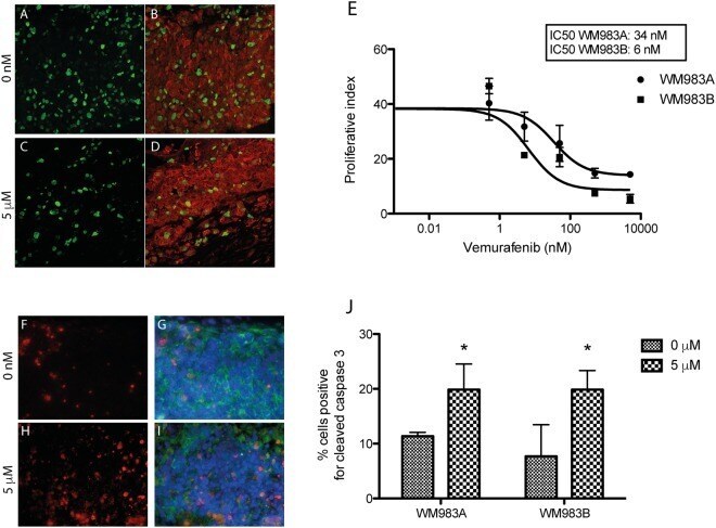
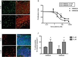
- Figure 7 Melanoma cell response to vemurafenib treatment in the 3D microenvironment. ( A - D ) Evaluation of melanoma cell proliferation in the model after 12 days of treatment with vemurafenib. Ki67 immunostaining (green signal) is indicative of cell proliferation in the WM983B melanoma tumor ( B , D , red signal, PMEL). ( E ) Proliferative index of WM983A and WM983B cells in the 3D model in response to increasing vemurafenib concentrations. ( F - I ) Co-labeling of apoptotic cells after staining for cleaved caspase 3 and tumor marker NG2. ( F , H ) Immunostaining for cleaved caspase 3 (red signal) in the melanoma model with ( G , I ) immunostaining of melanoma cells (neural/glial antigen 2 (NG-2), green signal and Hoechst, blue signal). ( J ) Quantification of cleaved caspase-3 expressing cells per total number of tumor cells as a function of vemurafenib concentration. (*p