 Explore
 Validate
 Learn
Learn Western blot
 Immunocytochemistry
Antibody data
- Antibody Data
- Antigen structure
- References [2]
- Comments [0]
- Validations
- Immunocytochemistry [1]
- Immunohistochemistry [1]
Submit
Validation data
Reference
Comment
Report error
- Product number
- PA1-16817 - Provider product page

- Provider
- Invitrogen Antibodies
- Product name
- NPC1 Polyclonal Antibody
- Antibody type
- Polyclonal
- Antigen
- Synthetic peptide
- Description
- By Western blot, PA1-16817 detects heterogeneously glycosylated NPC1 protein with prominent bands at ~170 and ~220 kDa. Suggested positive control: human fibroblast cell lysate.
- Reactivity
- Human, Mouse, Rat, Hamster, Porcine
- Host
- Rabbit
- Isotype
- IgG
- Vial size
- 100 μL
- Concentration
- 1 mg/mL
- Storage
- Store at 4°C short term. For long term storage, store at -20°C, avoiding freeze/thaw cycles.
Submitted references Tonic prime-boost of STING signalling mediates Niemann-Pick disease type C.
Multiple cationic amphiphiles induce a Niemann-Pick C phenotype and inhibit Ebola virus entry and infection.
Chu TT, Tu X, Yang K, Wu J, Repa JJ, Yan N
Nature 2021 Aug;596(7873):570-575
Nature 2021 Aug;596(7873):570-575
Multiple cationic amphiphiles induce a Niemann-Pick C phenotype and inhibit Ebola virus entry and infection.
Shoemaker CJ, Schornberg KL, Delos SE, Scully C, Pajouhesh H, Olinger GG, Johansen LM, White JM
PloS one 2013;8(2):e56265
PloS one 2013;8(2):e56265
No comments: Submit comment
Supportive validation
- Submitted by
- Invitrogen Antibodies (provider)
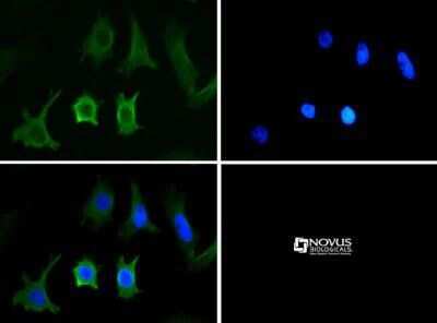
- Main image
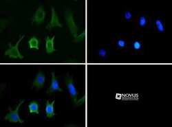
- Experimental details
- Immunocytochemistry analysis of NPC1 in HeLa cells. Samples were incubated in NPC1 polyclonal antibody (Product # PA1-16817) followed by DyLight 488 (green). Nuclei were counterstained with DAPI (blue).
Supportive validation
- Submitted by
- Invitrogen Antibodies (provider)
- Main image
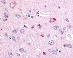
- Experimental details
- Immunohistochemical analysis of NPC1 in human brain, cortex, neurons and astrocytes. Samples were incubated in NPC1 polyclonal antibody (Product # PA1-16817).
