Explore
 Validate
 Learn
Learn Western blot
 Immunocytochemistry
Antibody data
- Antibody Data
- Antigen structure
- References [3]
- Comments [0]
- Validations
- Immunocytochemistry [1]
Submit
Validation data
Reference
Comment
Report error
- Product number
- HPA001473 - Provider product page

- Provider
- Atlas Antibodies
- Proper citation
- Atlas Antibodies Cat#HPA001473, RRID:AB_1078401
- Product name
- Anti-CASP9
- Antibody type
- Polyclonal
- Description
- Polyclonal Antibody against Human CASP9, Gene description: caspase 9, apoptosis-related cysteine peptidase, Alternative Gene Names: APAF-3, ICE-LAP6, MCH6, PPP1R56, Validated applications: WB, IHC, ICC, Uniprot ID: P55211, Storage: Store at +4°C for short term storage. Long time storage is recommended at -20°C.
- Reactivity
- Human, Mouse, Rat
- Host
- Rabbit
- Conjugate
- Unconjugated
- Isotype
- IgG
- Vial size
- 100 µl
- Concentration
- 0.05 mg/ml
- Storage
- Store at +4°C for short term storage. Long time storage is recommended at -20°C.
- Handling
- The antibody solution should be gently mixed before use.
Submitted references Mitochondrial levels determine variability in cell death by modulating apoptotic gene expression
CASP9 germline mutation in a family with multiple brain tumors
Placental Underperfusion in a Rat Model of Intrauterine Growth Restriction Induced by a Reduced Plasma Volume Expansion
Márquez-Jurado S, Díaz-Colunga J, das Neves R, Martinez-Lorente A, Almazán F, Guantes R, Iborra F
Nature Communications 2018;9(1)
Nature Communications 2018;9(1)
CASP9 germline mutation in a family with multiple brain tumors
Ronellenfitsch M, Oh J, Satomi K, Sumi K, Harter P, Steinbach J, Felsberg J, Capper D, Voegele C, Durand G, McKay J, Le Calvez‐Kelm F, Schittenhelm J, Klink B, Mittelbronn M, Ohgaki H
Brain Pathology 2017;28(1):94-102
Brain Pathology 2017;28(1):94-102
Placental Underperfusion in a Rat Model of Intrauterine Growth Restriction Induced by a Reduced Plasma Volume Expansion
Asselin E, Bibeau K, Sicotte B, Béland M, Bhat M, Gaboury L, Couture R, St-Louis J, Brochu M
PLOS ONE 2016;11(1):e0145982
PLOS ONE 2016;11(1):e0145982
No comments: Submit comment
Supportive validation
- Submitted by
- Atlas Antibodies (provider)
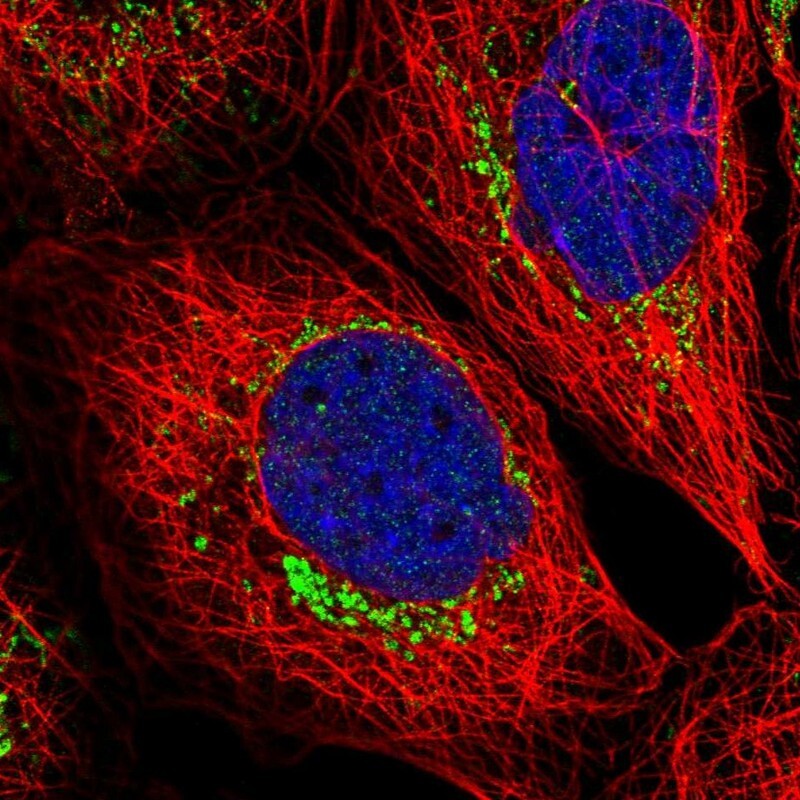
- Main image
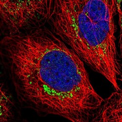
- Experimental details
- Immunofluorescent staining of human cell line A-431 shows localization to mitochondria.
- Sample type
- Human