Explore
 Validate
 Learn
Learn Western blot
 Other assay
Antibody data
- Antibody Data
- Antigen structure
- References [2]
- Comments [0]
- Validations
- Other assay [2]
Submit
Validation data
Reference
Comment
Report error
- Product number
- MA1-12562 - Provider product page

- Provider
- Invitrogen Antibodies
- Product name
- Caspase 9 Monoclonal Antibody (LAAP6 96 2-22)
- Antibody type
- Monoclonal
- Antigen
- Recombinant full-length protein
- Description
- A suggested positive control for this product is MCF-7 cell lysate.
- Reactivity
- Human
- Host
- Mouse
- Isotype
- IgG
- Antibody clone number
- LAAP6 96 2-22
- Vial size
- 100 μg
- Concentration
- 1 mg/mL
- Storage
- -20°C, Avoid Freeze/Thaw Cycles
Submitted references Identification of CETP as a molecular target for estrogen positive breast cancer cell death by cholesterol depleting agents.
Esculetin induces antiproliferative and apoptotic response in pancreatic cancer cells by directly binding to KEAP1.
Esau L, Sagar S, Bangarusamy D, Kaur M
Genes & cancer 2016 Sep;7(9-10):309-322
Genes & cancer 2016 Sep;7(9-10):309-322
Esculetin induces antiproliferative and apoptotic response in pancreatic cancer cells by directly binding to KEAP1.
Arora R, Sawney S, Saini V, Steffi C, Tiwari M, Saluja D
Molecular cancer 2016 Oct 18;15(1):64
Molecular cancer 2016 Oct 18;15(1):64
No comments: Submit comment
Supportive validation
- Submitted by
- Invitrogen Antibodies (provider)
- Main image
- Experimental details
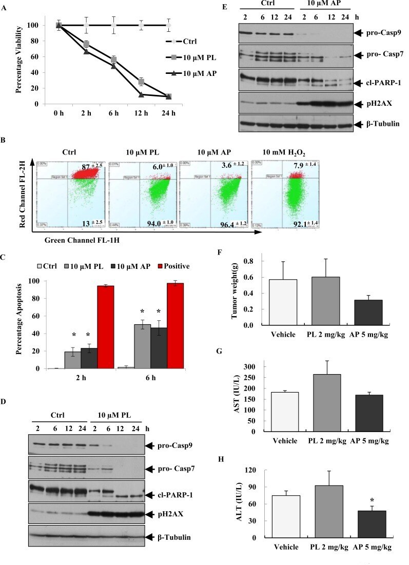
- Figure 1 AP induces intrinsic apoptosis in MCF-7 cells and reduces tumour burden in MCF-7 xenograft models (A) Viability of MCF-7 cells over time after treatment with 10 muM PL and AP. (B) MOMP disruption as measured by flow cytometry after treating MCF-7 cells with 10 muM PL and AP for 1 h; Ctrl represents cells treated with 0.2% DMSO. (C) The apoptosis-inducing potential of 10 muM PL and AP in MCF-7 as determined by using APOPercentage assay performed at 2 and 6 h. Data shown (in A, B, and C) are representative of mean +- SD of quadruplicate wells/condition in at least two independent experiments (n=2).* Indicates P
- Submitted by
- Invitrogen Antibodies (provider)
- Main image

- Experimental details
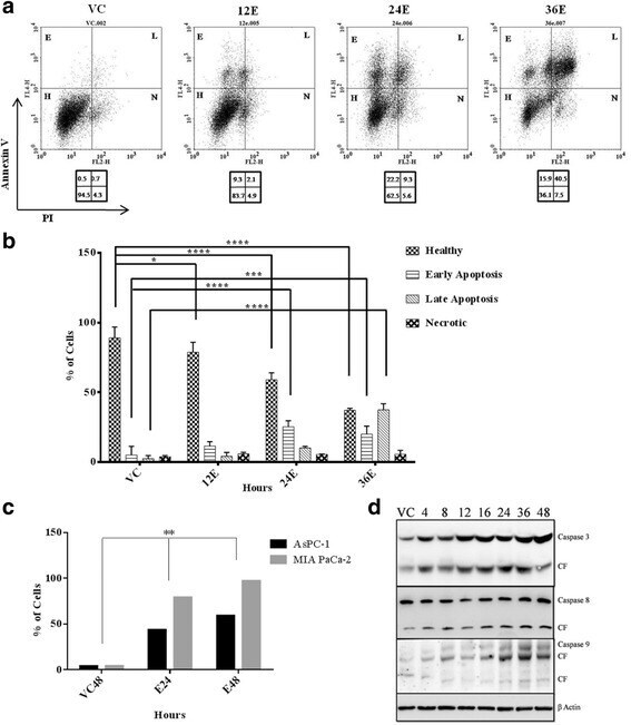
- Fig. 2 Esculetin induces apoptosis in pancreatic cancer cells: a Flow cytometric analysis of PANC-1 cells treated with 100 muM esculetin for indicated time showed temporal increase in surface expression of apoptotic marker- Annexin V indicating increased population of cells in apoptotic phase. b Percentage of PANC-1 cells exhibiting fluorescence in all four panels (healthy, early apoptosis, late apoptosis and necrosis) showing time dependent increase in apoptosis in Ecsuletin treated cells. c Percentage of cells with active APO BrdU indicating apoptosis in the absence and presence of esculetin (100 muM) as determined using TUNEL assay. d Western blot analysis showing an increase in expression of pro and active form of caspases (VC stands for vehicle control, E stands for esculetin treated sample for indicated time, CF stands for cleaved form, numerals represent time of esculetin treatment). Data represents the mean +- SD of three independent experiments. The significance was determined using ANOVA (Bonferroni's test). Key:* p < 0.05; ** p < 0.01; *** p < 0.001; **** p < 0.0001)