Explore
 Validate
 Learn
Learn Western blot
 Immunocytochemistry
 Immunohistochemistry
Antibody data
- Antibody Data
- Antigen structure
- References [4]
- Comments [0]
- Validations
- Immunocytochemistry [2]
- Other assay [1]
Submit
Validation data
Reference
Comment
Report error
- Product number
- MA1-16842 - Provider product page

- Provider
- Invitrogen Antibodies
- Product name
- Caspase 9 Monoclonal Antibody (LAP6 96-2-22)
- Antibody type
- Monoclonal
- Antigen
- Recombinant full-length protein
- Description
- Suggested positive control: HEK 293 whole cell lysate.
- Reactivity
- Human
- Host
- Mouse
- Isotype
- IgG
- Antibody clone number
- LAP6 96-2-22
- Vial size
- 200 μL
- Concentration
- 1 mg/mL
- Storage
- -20°C, Avoid Freeze/Thaw Cycles
Submitted references Simple and Efficient Protocol for Subcellular Fractionation of Normal and Apoptotic Cells.
The expressions of HSP70 and αB-crystallin in myocarditis associated with foot-and-mouth disease virus in lambs.
The expressions of HSP70 and αB-crystallin in myocarditis associated with foot-and-mouth disease virus in lambs.
Ovarian expression of markers associated with proliferation or apoptosis in women with diminished ovarian reserve.
Senichkin VV, Prokhorova EA, Zhivotovsky B, Kopeina GS
Cells 2021 Apr 9;10(4)
Cells 2021 Apr 9;10(4)
The expressions of HSP70 and αB-crystallin in myocarditis associated with foot-and-mouth disease virus in lambs.
Gulbahar MY, Kabak YB, Karayigit MO, Yarim M, Guvenc T, Parlak U
Journal of veterinary science 2011 Mar;12(1):65-73
Journal of veterinary science 2011 Mar;12(1):65-73
The expressions of HSP70 and αB-crystallin in myocarditis associated with foot-and-mouth disease virus in lambs.
Gulbahar MY, Kabak YB, Karayigit MO, Yarim M, Guvenc T, Parlak U
Journal of veterinary science 2011 Mar;12(1):65-73
Journal of veterinary science 2011 Mar;12(1):65-73
Ovarian expression of markers associated with proliferation or apoptosis in women with diminished ovarian reserve.
Vital-Reyes V, Rodríguez-Burford C, Chhieng DC, Alvarado-Cabrero I, Reyes-Fuentes A, Grizzle WE
Fertility and sterility 2006 Jul;86(1):176-85
Fertility and sterility 2006 Jul;86(1):176-85
No comments: Submit comment
Supportive validation
- Submitted by
- Invitrogen Antibodies (provider)
- Main image
- Experimental details
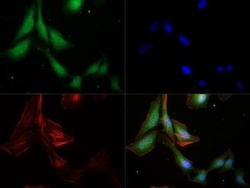
- Immunocytochemistry analysis of Caspase 9 in HeLa cells. Samples were incubated in Caspase 9 monoclonal antibody (Product # MA1-16842) followed by DyLight 488 (Green). Alpha-tubulin and nuclei were counterstained against DyLight 550 (Red) and DAPI (Blue).

- Submitted by
- Invitrogen Antibodies (provider)
- Main image

- Experimental details
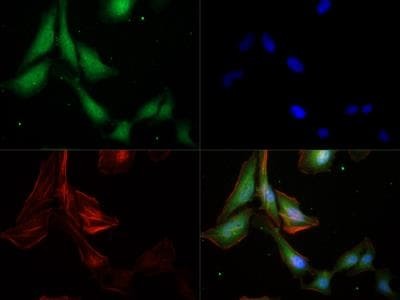
- Immunocytochemistry analysis of Caspase 9 in HeLa cells. Samples were incubated in Caspase 9 monoclonal antibody (Product # MA1-16842) followed by DyLight 488 (Green). Alpha-tubulin and nuclei were counterstained against DyLight 550 (Red) and DAPI (Blue).
Supportive validation
- Submitted by
- Invitrogen Antibodies (provider)
- Main image
- Experimental details
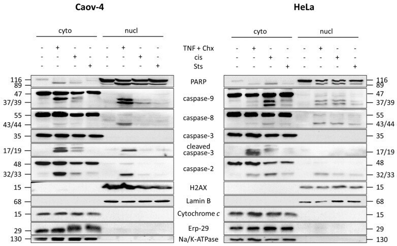
- Figure 3 Validation of the L&W protocol for nucleus/cytoplasm fractionation of apoptotic cells. WB analysis of cytoplasmic and nuclear fractions from Caov-4 and HeLa cells treated with 0.1 uM staurosporine (Sts), 10 ng/mL TNF-alpha + 5 mug/mL cycloheximide (TNF + Chx), or 35 uM cisplatin (cis). The purity of the resulting fractions was assessed by staining for markers of the cell membrane (Na/K ATPase), ER (ERp-29), mitochondria (cytochrome c ), nuclear envelope (Lamin B), and nucleoplasm (H2AX). PARP cleavage was evaluated as a marker of cell death. Cyto, cytoplasmic fraction; nucl, nuclear fraction.