Explore
 Validate
 Learn
Learn Western blot
 Immunocytochemistry
Antibody data
- Antibody Data
- Antigen structure
- References [6]
- Comments [0]
- Validations
- Immunocytochemistry [6]
- Other assay [5]
Submit
Validation data
Reference
Comment
Report error
- Product number
- PA5-17913 - Provider product page

- Provider
- Invitrogen Antibodies
- Product name
- Caspase 9 (Cleaved Asp315) Polyclonal Antibody
- Antibody type
- Polyclonal
- Antigen
- Synthetic peptide
- Description
- It is not recommended to aliquot this antibody. This antibody was orginally validated as part of a Thermo Scientific Cellomics High Content Screening Kit. The antibody sold separately may have slightly different performance and may need to be further optimized for the best results.
- Reactivity
- Human
- Host
- Rabbit
- Isotype
- IgG
- Vial size
- 100 μL
- Concentration
- 6 μg/mL
- Storage
- -20°C
Submitted references Purine nucleoside phosphorylase deficiency induces p53-mediated intrinsic apoptosis in human induced pluripotent stem cell-derived neurons.
miR‑302d‑3p regulates the viability, migration and apoptosis of breast cancer cells through regulating the TMBIM6‑mediated ERK signaling pathway.
Stingray Venom Proteins: Mechanisms of Action Revealed Using a Novel Network Pharmacology Approach.
Inhibition of the prolyl isomerase Pin1 enhances the ability of sorafenib to induce cell death and inhibit tumor growth in hepatocellular carcinoma.
Long Non-Coding RNA (lncRNA) Urothelial Carcinoma-Associated 1 (UCA1) Enhances Tamoxifen Resistance in Breast Cancer Cells via Inhibiting mTOR Signaling Pathway.
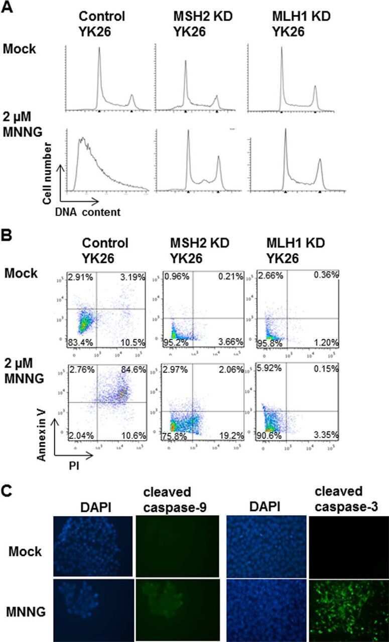
Human pluripotent stem cells have a novel mismatch repair-dependent damage response.
Tsui M, Biro J, Chan J, Min W, Dobbs K, Notarangelo LD, Grunebaum E
Scientific reports 2022 May 31;12(1):9084
Scientific reports 2022 May 31;12(1):9084
miR‑302d‑3p regulates the viability, migration and apoptosis of breast cancer cells through regulating the TMBIM6‑mediated ERK signaling pathway.
Liao Y, Qiu Z, Bai L
Molecular medicine reports 2021 Dec;24(6)
Molecular medicine reports 2021 Dec;24(6)
Stingray Venom Proteins: Mechanisms of Action Revealed Using a Novel Network Pharmacology Approach.
Kirchhoff KN, Billion A, Voolstra CR, Kremb S, Wilke T, Vilcinskas A
Marine drugs 2021 Dec 24;20(1)
Marine drugs 2021 Dec 24;20(1)
Inhibition of the prolyl isomerase Pin1 enhances the ability of sorafenib to induce cell death and inhibit tumor growth in hepatocellular carcinoma.
Zheng M, Xu H, Liao XH, Chen CP, Zhang AL, Lu W, Wang L, Yang D, Wang J, Liu H, Zhou XZ, Lu KP
Oncotarget 2017 May 2;8(18):29771-29784
Oncotarget 2017 May 2;8(18):29771-29784
Long Non-Coding RNA (lncRNA) Urothelial Carcinoma-Associated 1 (UCA1) Enhances Tamoxifen Resistance in Breast Cancer Cells via Inhibiting mTOR Signaling Pathway.
Wu C, Luo J
Medical science monitor : international medical journal of experimental and clinical research 2016 Oct 21;22:3860-3867
Medical science monitor : international medical journal of experimental and clinical research 2016 Oct 21;22:3860-3867
Human pluripotent stem cells have a novel mismatch repair-dependent damage response.
Lin B, Gupta D, Heinen CD
The Journal of biological chemistry 2014 Aug 29;289(35):24314-24
The Journal of biological chemistry 2014 Aug 29;289(35):24314-24
No comments: Submit comment
Supportive validation
- Submitted by
- Invitrogen Antibodies (provider)
- Main image
- Experimental details
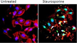
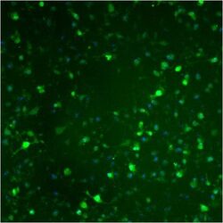
- Immunofluorescence staining of cleaved (active) caspase 9 in HeLa cells treated with vehicle (0.1% DMSO in media) or with 1 µM staurosporine for 4 hours. Cells were stained according to the kit protocol and imaged using a Cellomics ArrayScan HCS Reader. Cells treated with stautosporine results in caspase 9 activation and an increase in staining.
- Submitted by
- Invitrogen Antibodies (provider)
- Main image
- Experimental details
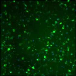
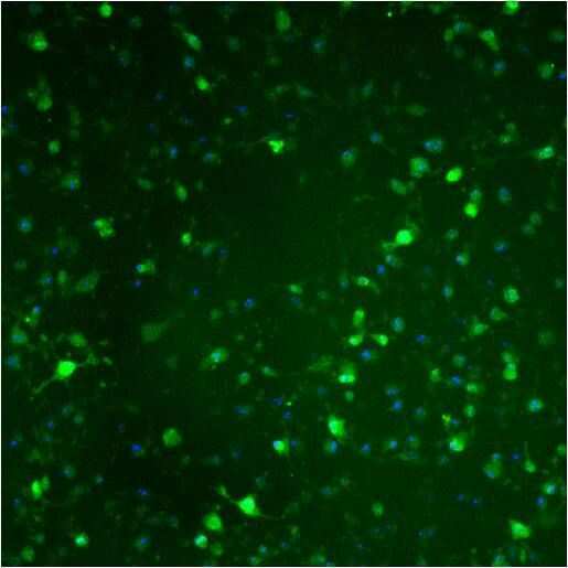
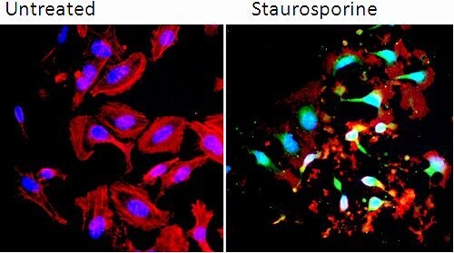
- Immunofluorescent analysis of Cleaved Caspase-9 (green) in HeLa cells either left untreated (left lane) or treated with 1µM Staurosporine for 3 hours. Formalin fixed cells were permeabilized with 0.1% Triton X-100 in TBS for 10 minutes at room temperature and blocked with 1% Blocker BSA (Product # 37525) for 15 minutes at room temperature. Cells were probed with a Cleaved Caspase-9 polyclonal antibody (Product # PA5-17913) at a dilution of 1:100 for at least 1 hour at room temperature, washed with PBS, and incubated with DyLight 488 goat anti-rabbit IgG secondary antibody (Product # 35552) at a dilution of 1:400 for 30 minutes at room temperature. F-Actin (red) was stained with Dylight 554 Phalloidin (Product # 21834) and nuclei (blue) were stained with Hoechst 33342 dye (Product # 62249). Images were taken on a Thermo Scientific ArrayScan or a ToxInsight Instrument at 20X magnification.

- Submitted by
- Invitrogen Antibodies (provider)
- Main image

- Experimental details
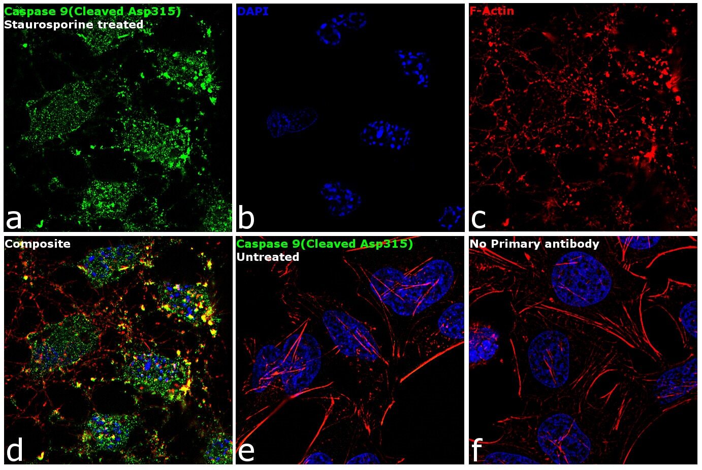
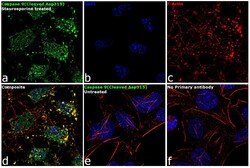
- Immunofluorescence analysis of Caspase 9 (Cleaved Asp315) was performed using 70% confluent log phase HeLa cells treated with 1µM of Staurosporine for 3 hours. The cells were fixed with 4% paraformaldehyde for 10 minutes, permeabilized with 0.1% Triton™ X-100 for 15 minutes, and blocked with 1% BSA for 1 hour at room temperature. The cells were labeled with Caspase 9 (Cleaved Asp315) Rabbit Polyclonal Antibody (Product # PA5-17913) at 1:100 dilution in 0.1% BSA, incubated at 4 degree Celsius overnight and then labeled with Goat anti-Rabbit IgG (H+L) Superclonal™ Secondary Antibody, Alexa Fluor® 488 conjugate (Product # A27034) at a dilution of 1:2000 for 45 minutes at room temperature (Panel a: green). Nuclei (Panel b: blue) were stained with SlowFade® Gold Antifade Mountant with DAPI (Product # S36938). F-actin (Panel c: red) was stained with Rhodamine Phalloidin (Product # R415, 1:300). Panel d represents the merged image showing cytoplasmic and nuclear localization. Panel e shows untreated cells with no signal. Panel f represents control cells with no primary antibody to assess background. The images were captured at 60X magnification.
- Submitted by
- Invitrogen Antibodies (provider)
- Main image

- Experimental details
- Immunofluorescent analysis of Cleaved Caspase-9 (green) in HeLa cells either left untreated (left lane) or treated with 1µM Staurosporine for 3 hours. Formalin fixed cells were permeabilized with 0.1% Triton X-100 in TBS for 10 minutes at room temperature and blocked with 1% Blocker BSA (Product # 37525) for 15 minutes at room temperature. Cells were probed with a Cleaved Caspase-9 polyclonal antibody (Product # PA5-17913) at a dilution of 1:100 for at least 1 hour at room temperature, washed with PBS, and incubated with DyLight 488 goat anti-rabbit IgG secondary antibody (Product # 35552) at a dilution of 1:400 for 30 minutes at room temperature. F-Actin (red) was stained with Dylight 554 Phalloidin (Product # 21834) and nuclei (blue) were stained with Hoechst 33342 dye (Product # 62249). Images were taken on a Thermo Scientific ArrayScan or a ToxInsight Instrument at 20X magnification.
- Submitted by
- Invitrogen Antibodies (provider)
- Main image
- Experimental details
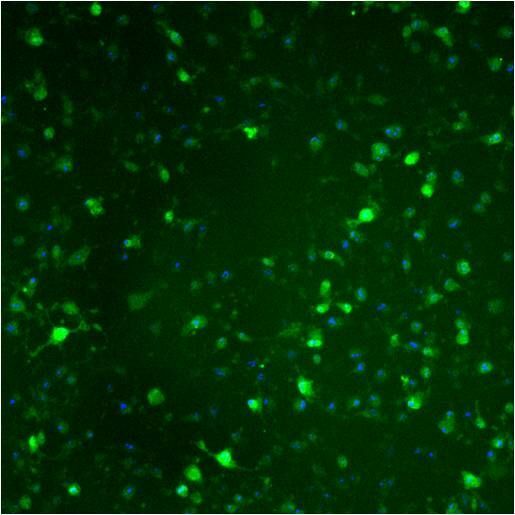
- Immunofluorescence staining of cleaved (active) caspase 9 in HeLa cells treated with vehicle (0.1% DMSO in media) or with 1 µM staurosporine for 4 hours. Cells were stained according to the kit protocol and imaged using a Cellomics ArrayScan HCS Reader. Cells treated with stautosporine results in caspase 9 activation and an increase in staining.
- Submitted by
- Invitrogen Antibodies (provider)
- Main image
- Experimental details
- Immunofluorescence analysis of Caspase 9 (Cleaved Asp315) was performed using 70% confluent log phase HeLa cells treated with 1µM of Staurosporine for 3 hours. The cells were fixed with 4% paraformaldehyde for 10 minutes, permeabilized with 0.1% Triton™ X-100 for 15 minutes, and blocked with 1% BSA for 1 hour at room temperature. The cells were labeled with Caspase 9 (Cleaved Asp315) Rabbit Polyclonal Antibody (Product # PA5-17913) at 1:100 dilution in 0.1% BSA, incubated at 4 degree Celsius overnight and then labeled with Goat anti-Rabbit IgG (Heavy Chain) Superclonal™ Secondary Antibody, Alexa Fluor® 488 conjugate (Product # A27034) at a dilution of 1:2000 for 45 minutes at room temperature (Panel a: green). Nuclei (Panel b: blue) were stained with SlowFade® Gold Antifade Mountant with DAPI (Product # S36938). F-actin (Panel c: red) was stained with Rhodamine Phalloidin (Product # R415, 1:300). Panel d represents the merged image showing cytoplasmic and nuclear localization. Panel e shows untreated cells with no signal. Panel f represents control cells with no primary antibody to assess background. The images were captured at 60X magnification.

Supportive validation
- Submitted by
- Invitrogen Antibodies (provider)
- Main image
- Experimental details
- NULL
- Submitted by
- Invitrogen Antibodies (provider)
- Main image
- Experimental details
- Figure 2 UCA1 knockdown sensitize breast cancer cells to tamoxifen. ( A, B ) CCK-8 assay of cell viability of LCC2 ( A ) and LCC9 ( B ) cells with or without UCA1 knockdown after treatment with varying concentrations of tamoxifen (0.1, 0.5, 1, 5, 10, 15, 20, and 50 mumol/L) for three days. ( C ) Typical images of the cleaved caspase-9 labeled by Alexa Fluor-555-labeled antibody (red color) and the nuclei stained by DAPI (blue color). ( D, E ) Representative images ( D ) and quantitation ( E ) of flow cytometric analysis of apoptotic LCC2 and LCC9 cells with or without UCA1 knockdown after treatment with 10 muM of tamoxifen for three days. * p

- Submitted by
- Invitrogen Antibodies (provider)
- Main image
- Experimental details
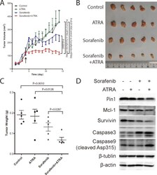
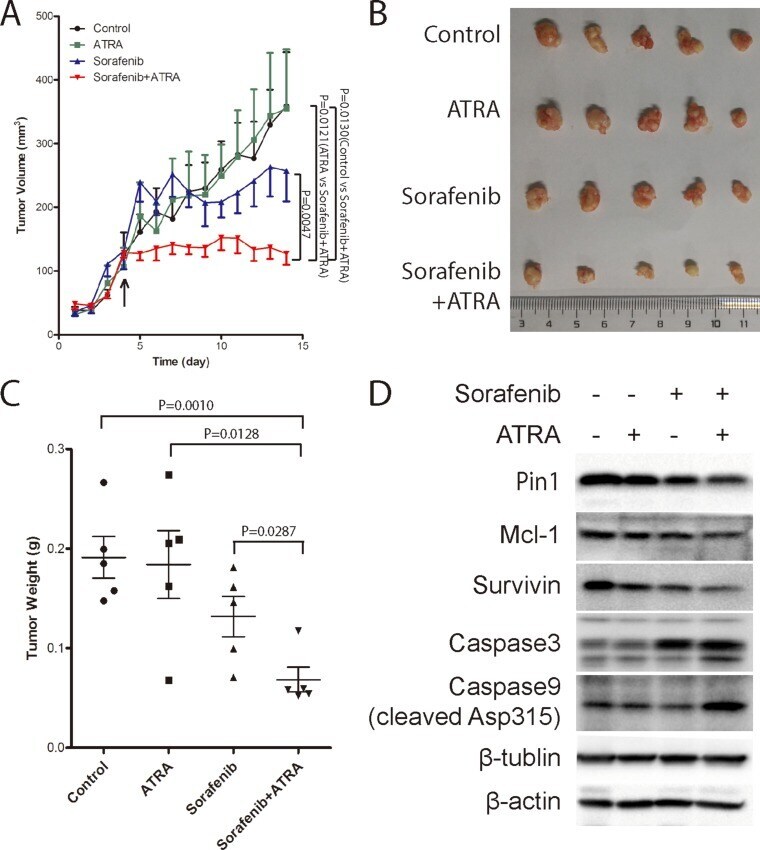
- Figure 7 The Pin1 inhibitor ATRA synergistically enhances the ability of sorafenib to inhibit tumor growth of HCC in vivo ( A ) Tumor growth was significantly inhibited by the combination of sorafenib and ATRA. Huh7 cells were injected subcutaneously into nude mice. Drugs were administrated when tumors at the point indicated by the arrow. 1/4 tablet of 10 mg 21 day slow-releasing ATRA pellet was inoculated subcutaneously. Sorafenib (40 mg/kg) was orally given every three days. Tumor growth was measured every 3 days, and tumor volume was calculated using the formula of length*width*width/2. ( B , C ) ATRA synergistically enhanced sorafenib anti-tumor effect in vivo . Tumors were harvested (B) and weighted (C) when the length of largest tumor reached 1.0 cm. ( D ) ATRA synergistically enhanced sorafenib induced Pin1 down-regulation in vivo . Pin1, Mcl-1, survivin, beta-actin, beta-tubulin, cleaved caspase 9 and caspase 3, protein expressions were determined by Western Blot.
- Submitted by
- Invitrogen Antibodies (provider)
- Main image
- Experimental details
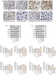
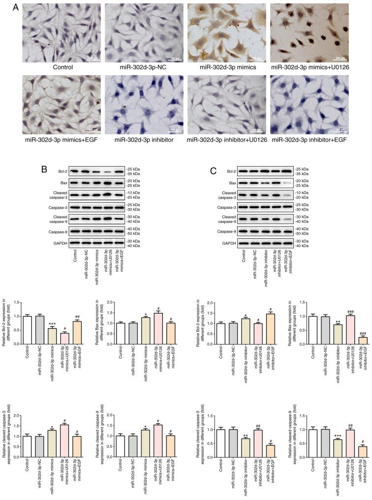
- Figure 9. miR-302d-3p regulates ERK signaling pathway by targeting transmembrane Bax inhibitor motif containing 6 to promote apoptosis of BC cells. (A) TUNEL assay was performed to detect the apoptosis rate of BC cells (scale bar, 100 um). Western blotting was performed to measure the expression of apoptosis-related proteins following transfection with (B) miR-302d-3p mimics or (C) miR-302d-3p inhibitor plus U0126 or EGF. *P
- Submitted by
- Invitrogen Antibodies (provider)
- Main image

- Experimental details
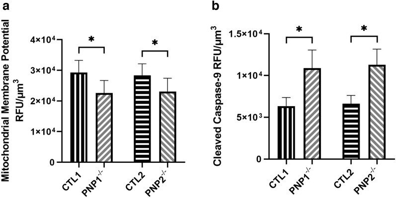
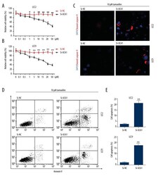
- Elevated intrinsic apoptosis in PNP-deficient induced pluripotent stem cell-derived neurons. The mitochondrial membrane potential ( A ) and cleaved caspase-9 ( B ) fluorescence of neurons derived from control (CTL) and PNP-deficient (PNP -/- ) iPSCs. The average relative fluorescence units (RFU)/um 3 was calculated by measuring fluorescence throughout the whole volume of a neuron. Data shown as mean + SD of n = 1050 (350 neurons from 3 replicates); * p < 0.001.