 Explore
 Validate
 Learn
Learn Western blot
 Immunocytochemistry
 Immunoprecipitation
Antibody data
- Antibody Data
- Antigen structure
- References [1]
- Comments [0]
- Validations
- Immunocytochemistry [2]
Submit
Validation data
Reference
Comment
Report error
- Product number
- ALX-210-838-R100 - Provider product page

- Provider
- Enzo Life Sciences
- Proper citation
- Enzo Life Sciences Cat#ALX-210-838-R100, RRID:AB_2050956
- Product name
- Caspase-9 (human) polyclonal antibody
- Antibody type
- Polyclonal
- Antigen
- Recombinant full length protein
- Reactivity
- Human
- Host
- Rabbit
- Vial size
- 100 μl
- Storage
- -20°C
- Handling
- Avoid freeze/thaw cycles.
Submitted references The prevention of spontaneous apoptosis of follicular lymphoma B cells by a follicular dendritic cell line: involvement of caspase-3, caspase-8 and c-FLIP.
Goval JJ, Thielen C, Bourguignon C, Greimers R, Dejardin E, Choi YS, Boniver J, de Leval L
Haematologica 2008 Aug;93(8):1169-77
Haematologica 2008 Aug;93(8):1169-77
No comments: Submit comment
Supportive validation
- Submitted by
- Enzo Life Sciences (provider)
- Main image
- Experimental details
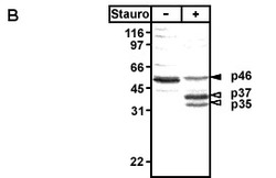
- (B) Detection of caspase-9 processing during apoptosis. The antiserum detects procaspase-9 (46kDa) and the intermediate cleavage products of 37kDa and 35 kDa. No cross-reactivity with other caspases isÊobserved. Method: Jurkat cells were treated with 2µM staurosporine (Prod. No. ALX-380-014). After 4 hours cell lysates were prepared and separated by SDS-Page (2x10E6 cells/lane) under reducing conditions. Proteins were immunoblotted with rabbit anti-caspase-9 (1:1'000). Following incubation with peroxidase-conjugated secondary antibodies caspase-9 processing was detected by enhanced chemoluminescent staining. The closed arrow indicates procaspase-9 (46kDa), which processed to the p35 and p37 fragments (open arrows).

- Submitted by
- Enzo Life Sciences (provider)
- Main image
- Experimental details
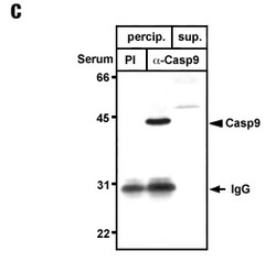
- (C) Immunoprecipitation of caspase-9. Method: Extracts of 1x10⁷ Jurkat cells were subjected to immunoprecipitation with Protein G-sepharose in the presence of rabbit preimmune serum (PI control, lane 1) or the antiserum to caspase-9 (lane 2). Lane 3 demonstrates that the antiserum is able to completely deplete caspase-9 from cell lysates, as no caspase-9 was detected in the supernatants after precipititaion.
