 Explore
 Validate
 Learn
Learn Western blot
 Immunohistochemistry
Antibody data
- Antibody Data
- Antigen structure
- References [6]
- Comments [0]
- Validations
- Western blot [3]
Submit
Validation data
Reference
Comment
Report error
- Product number
- MAB8301 - Provider product page

- Provider
- R&D Systems
- Product name
- Human Caspase-9 Antibody
- Antibody type
- Monoclonal
- Description
- Protein A or G purified from hybridoma culture supernatant. Detects human Caspase-9 in Western blots and captures Caspase-9 complexed with APAF-1.
- Reactivity
- Human
- Host
- Mouse
- Conjugate
- Unconjugated
- Isotype
- IgG
- Antibody clone number
- LAP6
- Vial size
- 100 ug
- Concentration
- LYOPH
- Storage
- Use a manual defrost freezer and avoid repeated freeze-thaw cycles. 12 months from date of receipt, -20 to -70 °C as supplied. 1 month, 2 to 8 °C under sterile conditions after reconstitution. 6 months, -20 to -70 °C under sterile conditions after reconstitution.
Submitted references Truncation of the TAR DNA-binding protein 43 is not a prerequisite for cytoplasmic relocalization, and is suppressed by caspase inhibition and by introduction of the A90V sequence variant.
Discovery of Sanggenon G as a natural cell-permeable small-molecular weight inhibitor of X-linked inhibitor of apoptosis protein (XIAP).
Intracellular K(+) inhibits apoptosis by suppressing the Apaf-1 apoptosome formation and subsequent downstream pathways but not cytochrome c release.
Hsp70 inhibits heat-induced apoptosis upstream of mitochondria by preventing Bax translocation.
X-linked inhibitor of apoptosis (XIAP) confers human trophoblast cell resistance to Fas-mediated apoptosis.
X-linked inhibitor of apoptosis (XIAP) confers human trophoblast cell resistance to Fas-mediated apoptosis.
Wobst HJ, Delsing L, Brandon NJ, Moss SJ
PloS one 2017;12(5):e0177181
PloS one 2017;12(5):e0177181
Discovery of Sanggenon G as a natural cell-permeable small-molecular weight inhibitor of X-linked inhibitor of apoptosis protein (XIAP).
Seiter MA, Salcher S, Rupp M, Hagenbuchner J, Kiechl-Kohlendorfer U, Mortier J, Wolber G, Rollinger JM, Obexer P, Ausserlechner MJ
FEBS open bio 2014;4:659-71
FEBS open bio 2014;4:659-71
Intracellular K(+) inhibits apoptosis by suppressing the Apaf-1 apoptosome formation and subsequent downstream pathways but not cytochrome c release.
Karki P, Seong C, Kim JE, Hur K, Shin SY, Lee JS, Cho B, Park IS
Cell death and differentiation 2007 Dec;14(12):2068-75
Cell death and differentiation 2007 Dec;14(12):2068-75
Hsp70 inhibits heat-induced apoptosis upstream of mitochondria by preventing Bax translocation.
Stankiewicz AR, Lachapelle G, Foo CP, Radicioni SM, Mosser DD
The Journal of biological chemistry 2005 Nov 18;280(46):38729-39
The Journal of biological chemistry 2005 Nov 18;280(46):38729-39
X-linked inhibitor of apoptosis (XIAP) confers human trophoblast cell resistance to Fas-mediated apoptosis.
Straszewski-Chavez SL, Abrahams VM, Funai EF, Mor G
Molecular human reproduction 2004 Jan;10(1):33-41
Molecular human reproduction 2004 Jan;10(1):33-41
X-linked inhibitor of apoptosis (XIAP) confers human trophoblast cell resistance to Fas-mediated apoptosis.
Straszewski-Chavez SL, Abrahams VM, Funai EF, Mor G
Molecular human reproduction 2004 Jan;10(1):33-41
Molecular human reproduction 2004 Jan;10(1):33-41
No comments: Submit comment
Supportive validation
- Submitted by
- R&D Systems (provider)
- Main image
- Experimental details
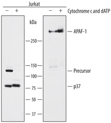
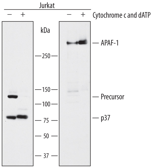
- Capture of Human Caspase-9 and Human Caspase-9 complexed with APAF-1 detected by Western Blot. Western blot shows Jurkat human acute T cell leukemia cell line lysates untreated (-) or treated (+) with 50 mM dATP and 1 mg/mL rat cytochrome c for 60 minutes, then captured on a 6-well dish coated at 10 µg/mL with Mouse Anti-Human Caspase-9 Monoclonal Antibody (Catalog # MAB8301). PVDF membrane was probed with 1 µg/mL of Mouse Anti-Human Caspase-9 Monoclonal Antibody (Catalog # MAB8301, left side) or Mouse Anti-Human APAF-1 Monoclonal Antibody (Catalog # MAB868, right side) followed by HRP-conjugated Anti-Mouse IgG Secondary Antibody (Catalog # HAF007). Specific bands were detected for Caspase-9 Precursor at approximately 46 kDa and the Caspase-9 p37 subunit at approximately 37 kDa (as indicated). A specific band was detected for APAF-1, captured as part of Caspase-9 complexed with APAF-1, at approximately 135 kDa (as indicated). This experiment was conducted under reducing conditions and using Immunoblot Buffer Group 4.
- Submitted by
- R&D Systems (provider)
- Main image
- Experimental details
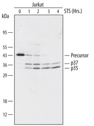
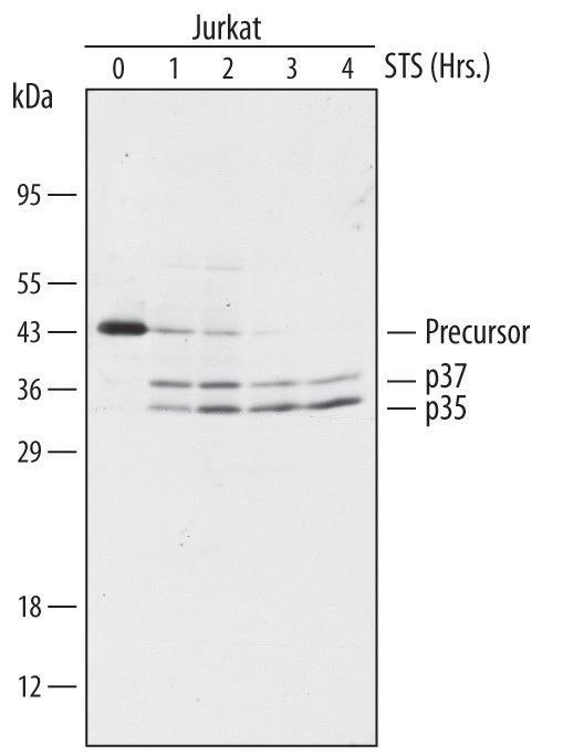
- Detection of Human Caspase-9 by Western Blot. Western blot shows lysates of Jurkat human acute T cell leukemia cell line treated with 1 µg/mL staurosporine (STS) for the indicated times. PVDF membrane was probed with 1 µg/mL of Mouse Anti-Human Caspase-9 Monoclonal Antibody (Catalog # MAB8301) followed by HRP-conjugated Anti-Mouse IgG Secondary Antibody (Catalog # HAF007). Specific bands were detected for Caspase-9 Precursor at approximately 46 kDa and the Caspase-9 p37 and p35 subunits at approximately 37 kDa and 35 kDa, respectively (as indicated) . This experiment was conducted under reducing conditions and using Immunoblot Buffer Group 4.
- Submitted by
- R&D Systems (provider)
- Main image
- Experimental details
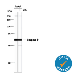
- Detection of Human Caspase-9 by Simple WesternTM. Simple Western lane view shows lysates of Jurkat human acute T cell leukemia cell line untreated (-) or treated (+) with 1 mM Staurosporine (STS) for 3 hours, loaded at 0.2 mg/mL. A specific band was detected for Caspase-9 at approximately 53 kDa (as indicated) using 20 µg/mL of Mouse Anti-Human Caspase-9 Monoclonal Antibody (Catalog # MAB8301). This experiment was conducted under reducing conditions and using the 12-230 kDa separation system. Non-specific interaction with the 230 kDa Simple Western standard may be seen with this antibody.
