Explore
 Validate
 Learn
Learn Western blot
Antibody data
- Antibody Data
- Antigen structure
- References [2]
- Comments [0]
- Validations
- Western blot [4]
- Immunoprecipitation [1]
- Immunohistochemistry [1]
Submit
Validation data
Reference
Comment
Report error
- Product number
- GTX100913 - Provider product page

- Provider
- GeneTex
- Proper citation
- GeneTex Cat#GTX100913, RRID:AB_2036363
- Product name
- B-Raf antibody [N2C1], Internal
- Antibody type
- Polyclonal
- Reactivity
- Human, Mouse, Rat
- Host
- Rabbit
Submitted references Acquired JHDM1D-BRAF Fusion Confers Resistance to FGFR Inhibition in FGFR2-Amplified Gastric Cancer.
Mitogen-Activated Protein Kinase Pathway: Genetic Analysis of 95 Adrenocortical Tumors.
Sase H, Nakanishi Y, Aida S, Horiguchi-Takei K, Akiyama N, Fujii T, Sakata K, Mio T, Aoki M, Ishii N
Molecular cancer therapeutics 2018 Oct;17(10):2217-2225
Molecular cancer therapeutics 2018 Oct;17(10):2217-2225
Mitogen-Activated Protein Kinase Pathway: Genetic Analysis of 95 Adrenocortical Tumors.
Rubin B, Monticelli H, Redaelli M, Mucignat C, Barollo S, Bertazza L, Mian C, Betterle C, Iacobone M, Fassina A, Boscaro M, Pezzani R, Mantero F
Cancer investigation 2015;33(10):526-31
Cancer investigation 2015;33(10):526-31
No comments: Submit comment
Supportive validation
- Submitted by
- GeneTex (provider)
- Main image

- Experimental details
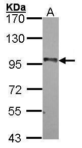
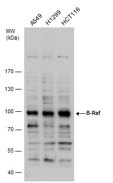
- Sample (30 ug of whole cell lysate) A: A549 7.5% SDS PAGE GTX100913 diluted at 1:1000
- Validation comment
- WB
- Submitted by
- GeneTex (provider)
- Main image
- Experimental details
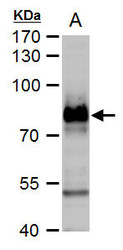
- B-Raf antibody [N2C1], Internal detects B-Raf protein by western blot analysis.A. 30 £gg PC-12 whole cell extract 7.5 % SDS-PAGEB-Raf antibody [N2C1], Internal (GTX100913) dilution: 1:5000
- Submitted by
- GeneTex (provider)
- Main image
- Experimental details
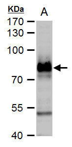
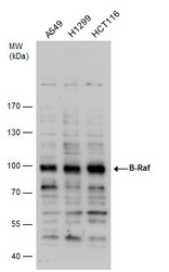
- B Raf antibody detects B Raf protein by western blot analysis. Various whole cell extracts (30 £gg) were separated by 7.5% SDS-PAGE, and the membrane was blotted with B Raf antibody (GTX100913) diluted by 1:1000.
- Submitted by
- GeneTex (provider)
- Main image
- Experimental details
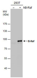
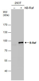
- B-Raf antibody [N2C1], Internal detects B-Raf protein by western blot analysis. Non-transfected (-) and B-Raf-transfected (+) 293T whole cell extracts (30 £gg) were separated by 7.5% SDS-PAGE, and the membrane was blotted with B-Raf antibody [N2C1], Internal (GTX100913) diluted at 1:5000.
Supportive validation
- Submitted by
- GeneTex (provider)
- Main image

- Experimental details
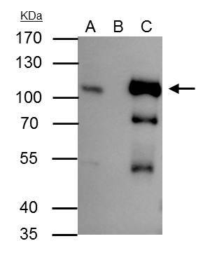
- B-Raf antibody [N2C1], Internal immunoprecipitates B-Raf protein in IP experiments. IP Sample: HepG2 whole cell lysate/extract A : 30 £gg whole cell lysate/extract of B-Raf protein expressing HepG2 cells B : Control with 3 £gg of pre-immune rabbit IgG C : Immunoprecipitation of B-Raf by 3 £gg of B-Raf antibody [N2C1], Internal (GTX100913) 10% SDS-PAGE The immunoprecipitated B-Raf protein was detected by B-Raf antibody [N2C1], Internal (GTX100913) diluted at 1 : 1000. EasyBlot anti-rabbit IgG (HRP) (GTX221666-01) was used as a secondary reagent.
Supportive validation
- Submitted by
- GeneTex (provider)
- Main image
- Experimental details
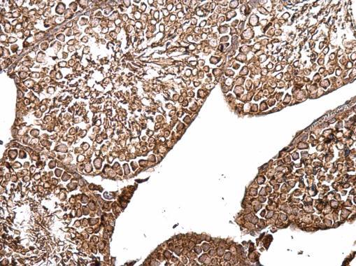
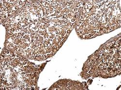
- B-Raf antibody [N2C1], Internal detects B-Raf protein at cytosol on mouse testis by immunohistochemical analysis. Sample: Paraffin-embedded mouse testis. B-Raf antibody [N2C1], Internal (GTX100913) dilution: 1:500.