Explore
 Validate
 Learn
Learn Western blot
Antibody data
- Antibody Data
- Antigen structure
- References [1]
- Comments [0]
- Validations
- Western blot [1]
- ELISA [1]
- Proximity ligation assay [1]
Submit
Validation data
Reference
Comment
Report error
- Product number
- H00000673-M02 - Provider product page

- Provider
- Abnova Corporation
- Proper citation
- Abnova Corporation Cat#H00000673-M02, RRID:AB_626391
- Product name
- BRAF monoclonal antibody (M02), clone 3D2
- Antibody type
- Monoclonal
- Description
- Mouse monoclonal antibody raised against a partial recombinant BRAF.
- Antigen sequence
FRPADEDHRNQFGQRDRSSSAPNVHINTIEPVNID
DLIRDQGFRGDGGSTTGLSATPPASLPGSLTNVKA
LQKSPGPQRERKSSSSSEDRNRMKTLGRRD- Isotype
- IgG
- Antibody clone number
- 3D2
- Storage
- Store at -20°C or lower. Aliquot to avoid repeated freezing and thawing.
Submitted references Analysis of protein-protein interactions in cross-talk pathways reveals CRKL protein as a novel prognostic marker in hepatocellular carcinoma.
Liu CH, Chen TC, Chau GY, Jan YH, Chen CH, Hsu CN, Lin KT, Juang YL, Lu PJ, Cheng HC, Chen MH, Chang CF, Ting YS, Kao CY, Hsiao M, Huang CY
Molecular & cellular proteomics : MCP 2013 May;12(5):1335-49
Molecular & cellular proteomics : MCP 2013 May;12(5):1335-49
No comments: Submit comment
Supportive validation
- Submitted by
- Abnova Corporation (provider)
- Main image
- Experimental details
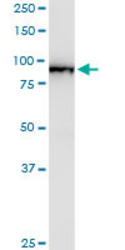
- BRAF monoclonal antibody (M02), clone 3D2. Western Blot analysis of BRAF expression in human pancreas.

Supportive validation
- Submitted by
- Abnova Corporation (provider)
- Main image
- Experimental details
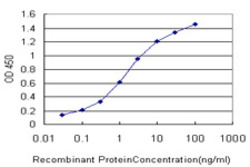
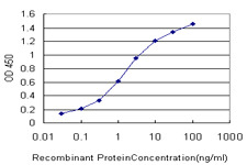
- Detection limit for recombinant GST tagged BRAF is approximately 0.03ng/ml as a capture antibody.
- Validation comment
- Sandwich ELISA (Recombinant protein)
- Protocol
- Protocol
Supportive validation
- Submitted by
- Abnova Corporation (provider)
- Main image

- Experimental details
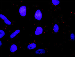
- Proximity Ligation Analysis of protein-protein interactions between MAPK3 and BRAF. HeLa cells were stained with anti-MAPK3 rabbit purified polyclonal 1:1200 and anti-BRAF mouse monoclonal antibody 1:50. Each red dot represents the detection of protein-protein interaction complex, and nuclei were counterstained with DAPI (blue).
- Validation comment
- In situ Proximity Ligation Assay (Cell)