Explore
 Validate
 Learn
Learn Western blot
Antibody data
- Antibody Data
- Antigen structure
- References [0]
- Comments [0]
- Validations
- Western blot [5]
- Immunocytochemistry [1]
Submit
Validation data
Reference
Comment
Report error
- Product number
- 711219 - Provider product page

- Provider
- Invitrogen Antibodies
- Product name
- B-Raf Recombinant Polyclonal Antibody (7HCLC)
- Antibody type
- Polyclonal
- Antigen
- Other
- Reactivity
- Human, Mouse
- Host
- Rabbit
- Isotype
- IgG
- Antibody clone number
- 7HCLC
- Vial size
- 100 µg
- Concentration
- 0.5 mg/mL
- Storage
- Store at 4°C short term. For long term storage, store at -20°C, avoiding freeze/thaw cycles.
No comments: Submit comment
Supportive validation
- Submitted by
- Invitrogen Antibodies (provider)
- Main image
- Experimental details
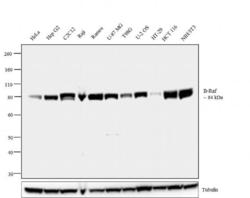
- Western blot analysis was performed on membrane enriched extracts (30 µg lysate) of HeLa (Lane 1), Hep G2 (Lane 2), C2C12 (Lane 3), Raji (Lane 4), Ramos (Lane 5), U-87 MG (Lane 6), T98G (Lane 7), U-2 OS (Lane 8), HT-29 (Lane 9), HCT116 (Lane 10) and NIH/3T3 (Lane 11). The blot was probed with Anti-B-Raf Recombinant Rabbit Polyclonal Antibody (Product # 711219, 1 µg/mL) and detected by chemiluminescence using Goat anti-Rabbit IgG (H+L) Superclonal™ Secondary Antibody, HRP conjugate (Product # A27036, 0.25 µg/mL, 1:4000 dilution). 84 kDa band corresponding to B-Raf was observed across the cell lines tested. Known quantity of protein samples were electrophoresed using Novex®NuPAGE®4-12 % Bis-Tris gel (Product # NP0321BOX), XCell SureLock™ Electrophoresis System (Product # EI0002) and Novex® Sharp Pre-Stained Protein Standard (Product # LC5800). Resolved proteins were then transferred onto a nitrocellulose membrane with iBlot® 2 Dry Blotting System (Product # IB21001). The membrane was probed with the relevant primary and secondary Antibody following blocking with 5 % skimmed milk. Chemiluminescent detection was performed using Pierce™ ECL Western blotting Substrate (Product # 32106).

- Submitted by
- Invitrogen Antibodies (provider)
- Main image
- Experimental details
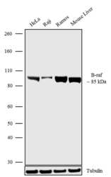
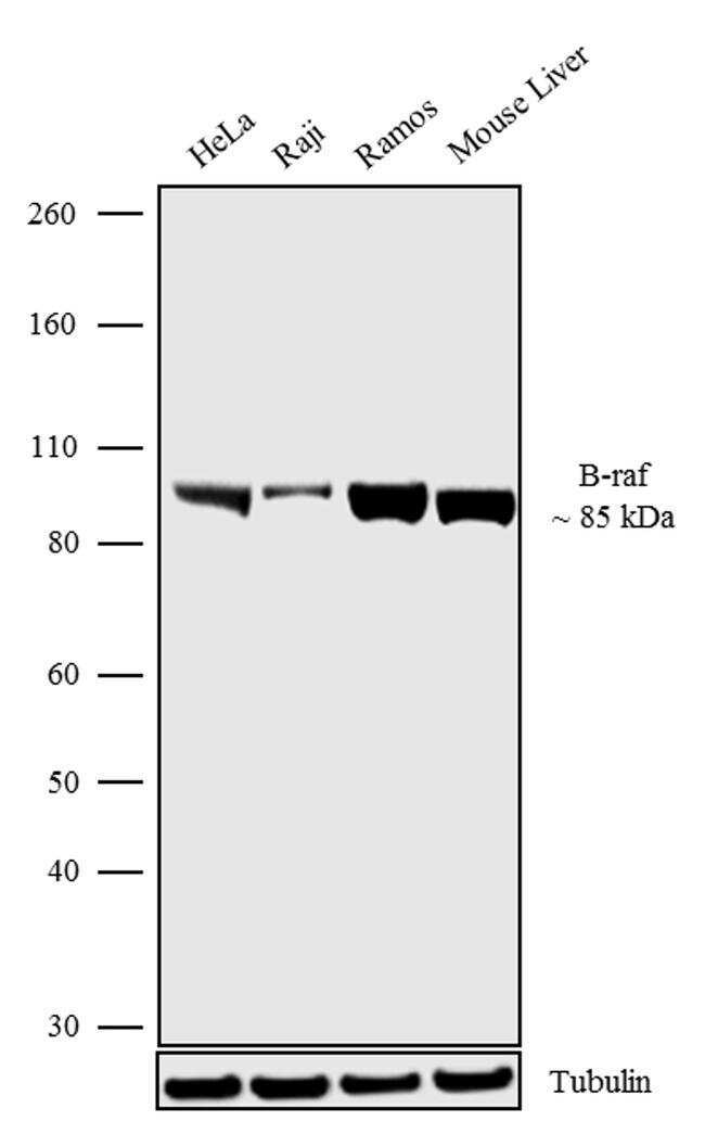
- Western blot analysis was performed on Whole cell extracts (30 µg lysate) of HeLa (Lane 1), Raji (Lane 2), Ramos (Lane 3) and tissue extracts of Mouse Liver (Lane 4). The blots were probed with Anti-B-raf Recombinant Rabbit Polyclonal Antibody (Product # 711219, 1-2 µg/mL) and detected by chemiluminescence using Goat anti-Rabbit IgG (H+L) Superclonal Secondary Antibody, HRP conjugate (Product # A27036, 0.4 µg/mL, 1:2500 dilution). A 85 kDa band corresponding to B-raf was observed across the cell lines and tissues tested. Known quantity of protein samples were electrophoresed using Novex®NuPAGE®4-12% Bis-Tris gel (Product # NP0321BOX), XCell SureLock Electrophoresis System (Product # EI0002) and Novex® Sharp Pre-Stained Protein Standard (Product # LC5800). Resolved proteins were then transferred onto a nitrocellulose membrane with iBlot® Dry Blotting System (Product # IB21001). The membrane was probed with the relevant primary and secondary Antibody following blocking with 5% skimmed milk. Chemiluminescent detection was performed using Pierce™ ECL Western blotting Substrate (Product # 32106).
- Submitted by
- Invitrogen Antibodies (provider)
- Main image

- Experimental details
- Western blot analysis was performed on membrane enriched extracts (30 µg lysate) of HeLa (Lane 1), Hep G2 (Lane 2), C2C12 (Lane 3), Raji (Lane 4), Ramos (Lane 5), U-87 MG (Lane 6), T98G (Lane 7), U-2 OS (Lane 8), HT-29 (Lane 9), HCT116 (Lane 10) and NIH/3T3 (Lane 11). The blot was probed with Anti-B-Raf Recombinant Rabbit Polyclonal Antibody (Product # 711219, 1 µg/mL) and detected by chemiluminescence using Goat anti-Rabbit IgG (H+L) Superclonal™ Secondary Antibody, HRP conjugate (Product # A27036, 0.25 µg/mL, 1:4000 dilution). 84 kDa band corresponding to B-Raf was observed across the cell lines tested. Known quantity of protein samples were electrophoresed using Novex®NuPAGE®4-12 % Bis-Tris gel (Product # NP0321BOX), XCell SureLock™ Electrophoresis System (Product # EI0002) and Novex® Sharp Pre-Stained Protein Standard (Product # LC5800). Resolved proteins were then transferred onto a nitrocellulose membrane with iBlot® 2 Dry Blotting System (Product # IB21001). The membrane was probed with the relevant primary and secondary Antibody following blocking with 5 % skimmed milk. Chemiluminescent detection was performed using Pierce™ ECL Western blotting Substrate (Product # 32106).

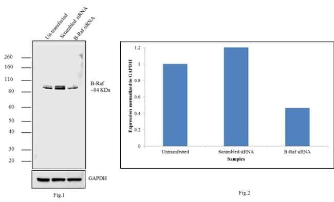
- Submitted by
- Invitrogen Antibodies (provider)
- Main image
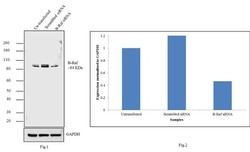
- Experimental details
- Knockdown of B-Raf was achieved by transfecting NIH/3T3 cells with B-Raf specific validated siRNAs (Silencer® select Product # s2080 + s2081 + s2082). Western blot analysis (Fig a) was performed using membrane enriched extracts from the B-Raf knockdown cells (lane 3), non-specific scrambled siRNA transfected cells (lane 2) and untransfected cells (lane 1). The blots were probed with Anti-B-Raf Recombinant Rabbit Polyclonal Antibody (Product # 711219, 1 µg/mL) and Goat anti-Rabbit IgG (H+L) Superclonal™ Secondary Antibody, HRP conjugate (Product # A27036, 0.25 µg/mL, 1:4000 dilution). Densitometric analysis of this Western blot is shown in histogram (Fig b). Decrease in signal upon siRNA mediated knock down confirms that antibody is specific to B-Raf.
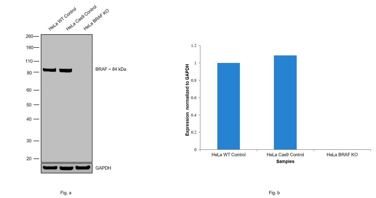
- Submitted by
- Invitrogen Antibodies (provider)
- Main image
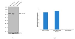
- Experimental details
- Knockout of BRAF was achieved by CRISPR-Cas9 genome editing using LentiArray™ Lentiviral sgRNA (Product # A32042) (Assay ID CRISPR868710_LV) and LentiArray Cas9 Lentivirus (Product # A32064). Western blot analysis of BRAF was performed by loading 30 µg of HeLa wild type (Lane 1), HeLa CAS9 (Lane 2), HeLa BRAF KO (Lane 3) whole cell extracts. The blot was probed with Anti-B-Raf Recombinant Polyclonal Antibody (7HCLC)(Product # 711219) using 1 µg/mL dilution and Goat anti-Rabbit IgG (H+L), Superclonal™ Recombinant Secondary Antibody, HRP (Product # A27036). Loss of signal upon CRISPR mediated knockout (KO) using the LentiArray™ CRISPR product line confirms that antibody is specific to BRAF.
Supportive validation
- Submitted by
- Invitrogen Antibodies (provider)
- Main image

- Experimental details
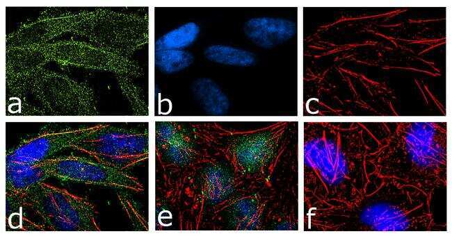
- For immunofluorescence analysis, HeLa cells were fixed and permeabilized for detection of Braf using Anti-Braf Recombinant Rabbit Polyclonal Antibody (Product # 711219, 2 µg/mL) and labeled with Goat anti-Rabbit IgG (H+L) Superclonal Secondary Antibody, Alexa Fluor® 488 conjugate (Product # A27034, 1:2000). Panel a) shows representative cells that were stained for detection and localization of Braf protein (green), Panel b) is stained for nuclei (blue) using SlowFade® Gold Antifade Mountant with DAPI (Product # S36938). Panel c) represents cytoskeletal F-actin staining using Alexa Fluor® 555 Rhodamine Phalloidin (Product # R415, 1:300). Panel d) is a composite image of Panels a, b and c clearly demonstrating cytoplasmic localization of Braf. Panel e) shows translocation of Braf into nucleus upon EGF treatment (100 ng/mL 5 minutes). Panel f) represents control cells with no primary antibody to assess background. The images were captured at 60X magnification.