Explore
 Validate
 Learn
Learn Western blot
 ELISA
ELISAAntibody data
- Antibody Data
- Antigen structure
- References [1]
- Comments [0]
- Validations
- Western blot [1]
- Immunocytochemistry [2]
- Immunohistochemistry [1]
- Other assay [1]
Submit
Validation data
Reference
Comment
Report error
- Product number
- MA5-15495 - Provider product page

- Provider
- Invitrogen Antibodies
- Product name
- B-Raf Monoclonal Antibody (1H12)
- Antibody type
- Monoclonal
- Antigen
- Purifed from natural sources
- Description
- MA5-15495 targets BRAF in indirect ELISA, IF, IHC, and WB applications and shows reactivity with Human and mouse samples. The MA5-15495 immunogen is purified recombinant fragment of human BRAF expressed in E. Coli.. MA5-15495 detects BRAF which has a predicted molecular weight of approximately 87kDa.
- Reactivity
- Human, Mouse
- Host
- Mouse
- Isotype
- IgG
- Antibody clone number
- 1H12
- Vial size
- 100 µL
- Concentration
- Conc. Not Determined
- Storage
- Store at 4°C short term. For long term storage, store at -20°C, avoiding freeze/thaw cycles.
Submitted references Oncogenic mutant RAS signaling activity is rescaled by the ERK/MAPK pathway.
Gillies TE, Pargett M, Silva JM, Teragawa CK, McCormick F, Albeck JG
Molecular systems biology 2020 Oct;16(10):e9518
Molecular systems biology 2020 Oct;16(10):e9518
No comments: Submit comment
Supportive validation
- Submitted by
- Invitrogen Antibodies (provider)
- Main image
- Experimental details
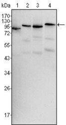
- Western blot analysis of BRAF using BRAF monoclonal antibody (Product # MA5-15495) in HeLa (1), HL60 (2), HepG2 (3) and NIH/3T3 (4) cell lysate.

Supportive validation
- Submitted by
- Invitrogen Antibodies (provider)
- Main image
- Experimental details
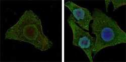
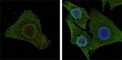
- Immunofluorescence analysis of MCF-7 (left) and HepG2 (right) cells using BRAF monoclonal antibody (Product # MA5-15495) (Green). Blue: DRAQ5 fluorescent DNA dye Red: Actin filaments have been labeled with DY-554 phalloidin.
- Submitted by
- Invitrogen Antibodies (provider)
- Main image
- Experimental details
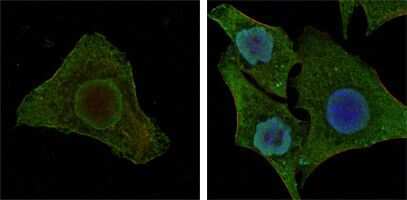
- Immunofluorescence analysis of MCF-7 (left) and HepG2 (right) cells using BRAF monoclonal antibody (Product # MA5-15495) (Green). Blue: DRAQ5 fluorescent DNA dye Red: Actin filaments have been labeled with DY-554 phalloidin.

Supportive validation
- Submitted by
- Invitrogen Antibodies (provider)
- Main image

- Experimental details
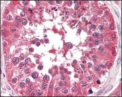
- Immunohistochemical analysis of paraffin-embedded human testis tissues using BRAF monoclonal antibody (Product # MA5-15495).
Supportive validation
- Submitted by
- Invitrogen Antibodies (provider)
- Main image

- Experimental details
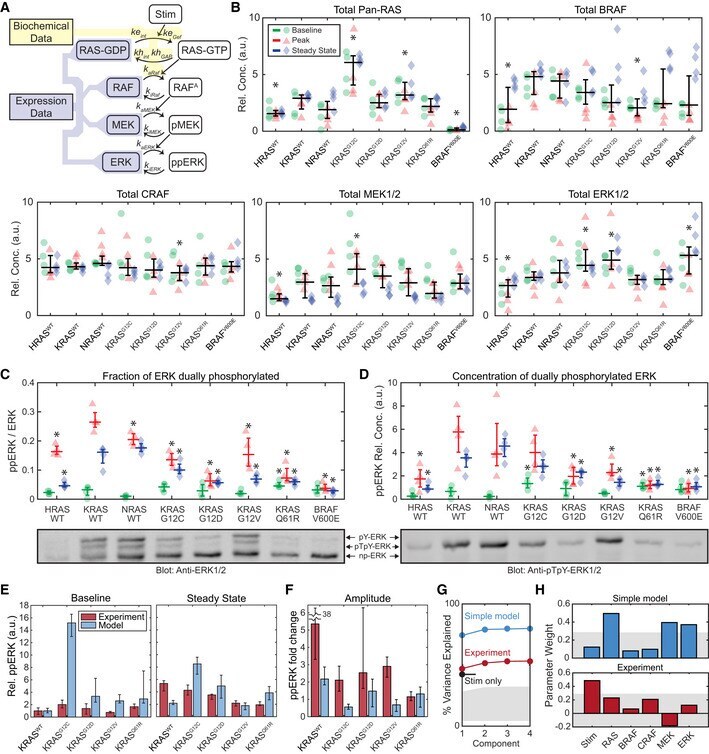
- Figure 5 Quantitative analysis of ERK phosphorylation in response to RAS mutation A Schematic of a model of the internal factors of the RAS-ERK pathway, showing parameters associated with each reaction. Shaded regions indicate portions of the model for which parameter values are available from either (yellow) published biochemical assays or (blue) our immunoblot expression data (B-D). B Immunoblot measurement of RAS-ERK pathway components in each cell line, at baseline (green circles), peak activity (15 min, red triangles), and steady-state activity (2 h, blue diamonds), four replicates each. Overlaid plots indicate the median (horizontal bars) and 25 th -75 th percentiles (vertical whiskers) over all conditions. Asterisks indicate statistical significance from the KRAS WT cell line, by t -test (pFDR < 0.05). C, D Phos-Tag immunoblot measurement of ERK fractional phosphorylation (C) and the relative concentration of dually-phosphorylated ERK (D), annotated as in (B), but with median and percentile ranges indicated per treatment condition, for 4 replicate cultures. Sample blot imagery shows anti-ERK1/2 (below C) and anti-ppERK1/2 (below D) for the same blot replicate. Asterisks indicate significance by t -test (pFDR < 0.05) from the KRAS WT cell line measurement for each condition. E, F Comparison of the internal RAS-ERK model to experimental data. (E) Relative ppERK as predicted by the internal model and measured by immunoblot for the 4 replicates collected, showing the media