Explore
 Validate
 Learn
Learn Western blot
 ELISA
ELISA Immunocytochemistry
Antibody data
- Antibody Data
- Antigen structure
- References [2]
- Comments [0]
- Validations
- Immunocytochemistry [2]
- Immunohistochemistry [3]
- Other assay [2]
Submit
Validation data
Reference
Comment
Report error
- Product number
- PA5-95830 - Provider product page

- Provider
- Invitrogen Antibodies
- Product name
- Decorin Polyclonal Antibody
- Antibody type
- Polyclonal
- Antigen
- Recombinant protein fragment
- Description
- Immunogen sequence: DEASGIGPEV PDDRDFEPSL GPVCPFRCQC HLRVVQCSDL GLDKVPKDLP PDTTLLDLQN NKITEIKDGD FKNLKNLHAL ILVNNKISKV SPGAFTPLVK LERLYLSKNQ LKELPEKMPK TLQELRAHEN EITKVRKVTF NGLNQMIVIE LGTNPLKSSG IENGAFQGMK KLSYIRIADT NITSIPQGLP PSLTELHLDG NKISRVDAAS LKGLNNLAKL GLSFNSISAV DNGSLANTPH LRELHLDNNK LTRVPGGLAE HKYIQVVYLH NNNISVVGSS DFCPPGHNTK KASYSGVSLF SNPVQYWEIQ PSTFRCVYVR SAIQLGNYK Positive Samples: Mouse heart, Mouse liver; Cellular Location: extracellular matrix, extracellular space
- Reactivity
- Human, Mouse, Rat
- Host
- Rabbit
- Isotype
- IgG
- Vial size
- 100 μL
- Concentration
- 0.88 mg/mL
- Storage
- -20°C, Avoid Freeze/Thaw Cycles
Submitted references Role of Decorin in Posterior Capsule Opacification and Eye Lens Development.
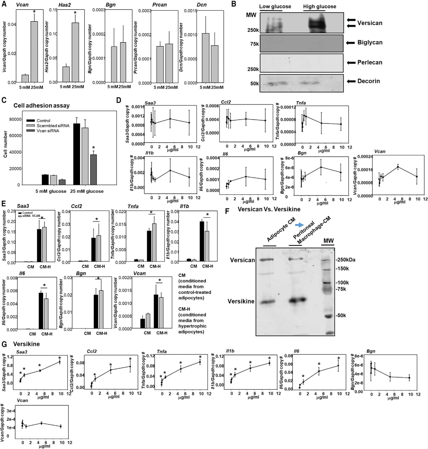
Adipocyte-Derived Versican and Macrophage-Derived Biglycan Control Adipose Tissue Inflammation in Obesity.
Shibata S, Shibata N, Ohtsuka S, Yoshitomi Y, Kiyokawa E, Yonekura H, Singh DP, Sasaki H, Kubo E
Cells 2021 Apr 9;10(4)
Cells 2021 Apr 9;10(4)
Adipocyte-Derived Versican and Macrophage-Derived Biglycan Control Adipose Tissue Inflammation in Obesity.
Han CY, Kang I, Harten IA, Gebe JA, Chan CK, Omer M, Alonge KM, den Hartigh LJ, Gomes Kjerulf D, Goodspeed L, Subramanian S, Wang S, Kim F, Birk DE, Wight TN, Chait A
Cell reports 2020 Jun 30;31(13):107818
Cell reports 2020 Jun 30;31(13):107818
No comments: Submit comment
Supportive validation
- Submitted by
- Invitrogen Antibodies (provider)
- Main image
- Experimental details
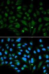
- Immunocytochemistry-Immunofluorescence analysis of Decorin was performed in MCF-7 cells using Decorin Polyclonal Antibody (Product # PA5-95830).

- Submitted by
- Invitrogen Antibodies (provider)
- Main image

- Experimental details
- Immunofluorescence analysis of Decorin in MCF-7 cells. Samples were incubated with Decorin Polyclonal antibody (Product # PA5-95830). Blue: DAPI for nuclear staining.

Supportive validation
- Submitted by
- Invitrogen Antibodies (provider)
- Main image

- Experimental details
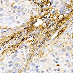
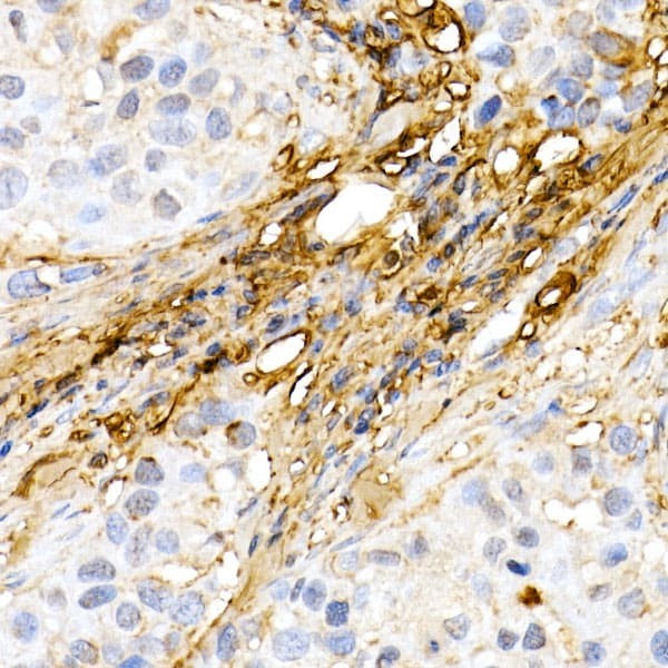
- Immunohistochemistry analysis of Decorin in paraffin-embedded human breast cancer using. Samples were incubated with Decorin Polyclonal antibody (Product # PA5-95830) using a dilution of 1:50 (40x lens). Perform high pressure antigen retrieval with 10 mM citrate buffer pH 6.0 before commencing with IHC staining protocol.

- Submitted by
- Invitrogen Antibodies (provider)
- Main image
- Experimental details
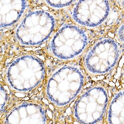
- Immunohistochemistry analysis of Decorin in paraffin-embedded human colon. Samples were incubated with Decorin Polyclonal antibody (Product # PA5-95830) using a dilution of 1:50 (40x lens). Perform high pressure antigen retrieval with 10 mM citrate buffer pH 6.0 before commencing with IHC staining protocol.

- Submitted by
- Invitrogen Antibodies (provider)
- Main image
- Experimental details
- Immunohistochemistry analysis of Decorin in paraffin-embedded human breast cancer using. Samples were incubated with Decorin Polyclonal antibody (Product # PA5-95830) using a dilution of 1:50 (40x lens). Perform high pressure antigen retrieval with 10 mM citrate buffer pH 6.0 before commencing with IHC staining protocol.
Supportive validation
- Submitted by
- Invitrogen Antibodies (provider)
- Main image
- Experimental details
- NULL
- Submitted by
- Invitrogen Antibodies (provider)
- Main image
- Experimental details
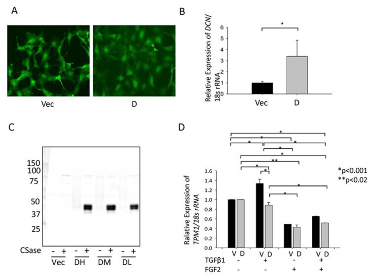
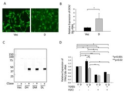
- Figure 4 Effect of overexpression of hDCN in HLECs. ( A ) We monitored the transduction efficiency of GFP-vector (Vec) and GFP-hDCN (D) by observing the GFP-expressing cells within the live cell population. ( B ) RT-qPCR was performed to confirm the expression level of hDCN mRNA in GFP-vector (Vec) and GFP-hDCN (D)-overexpressing SRA-HLECs. * p < 0.05. ( C ) The collected culture supernatant was subjected to SDS-PAGE. In total, 250 ng protein was loaded per lane after concentrating the culture supernatant and digesting the GAG chain with or without protease-free chondroitinase (CSase). The secretion of DCN from SRA-HLEC overexpressing GFP-hDCN (Expression level: DH, High; DM, moderate; DL, Low) or GFP-Vec (Vec) was confirmed using western blotting. The core protein of DCN was approximately 50 kDa. ( D ) Cultured GFP-vector (V) or GFP-hDCN (D)-transfected SRA-HLECs were plated in 35-mm dishes at the density of 8 x 10 4 in DMEM with 20% FBS for 24 h. LECs were treated with 10 ng/mL TGFbeta-2 and/or 100 ng/mL FGF2 in DMEM containing 1% FBS and incubated for 2 days. TPM1 mRNA level was analyzed using RT-qPCR. PC; Positive control, human dermis (1 mug/lane). * p < 0.001, ** p < 0.02.