Explore
 Validate
 Learn
Learn Western blot
 Immunoprecipitation
Antibody data
- Antibody Data
- Antigen structure
- References [3]
- Comments [0]
- Validations
- Western blot [2]
- Immunocytochemistry [1]
- Immunohistochemistry [2]
- Other assay [3]
Submit
Validation data
Reference
Comment
Report error
- Product number
- PA5-27370 - Provider product page

- Provider
- Invitrogen Antibodies
- Product name
- Decorin Polyclonal Antibody
- Antibody type
- Polyclonal
- Antigen
- Recombinant full-length protein
- Description
- Recommended positive controls: A549, H1299. Predicted reactivity: Mouse (82%), Rat (80%), Dog (91%), Pig (88%), Rabbit (90%), Sheep (86%), Chimpanzee (100%), Bovine (87%), Guinea pig (87%). Store product as a concentrated solution. Centrifuge briefly prior to opening the vial.
- Reactivity
- Human, Rat
- Host
- Rabbit
- Isotype
- IgG
- Vial size
- 100 μL
- Concentration
- 1 mg/mL
- Storage
- Store at 4°C short term. For long term storage, store at -20°C, avoiding freeze/thaw cycles.
Submitted references Capture and Detection of Circulating Glioma Cells Using the Recombinant VAR2CSA Malaria Protein.
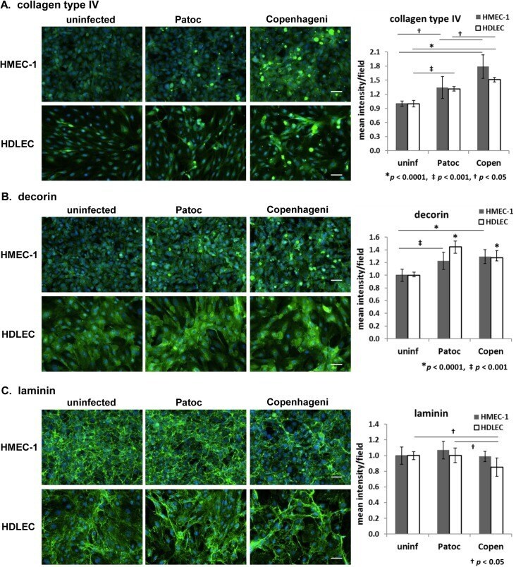
Leptospira interrogans causes quantitative and morphological disturbances in adherens junctions and other biological groups of proteins in human endothelial cells.
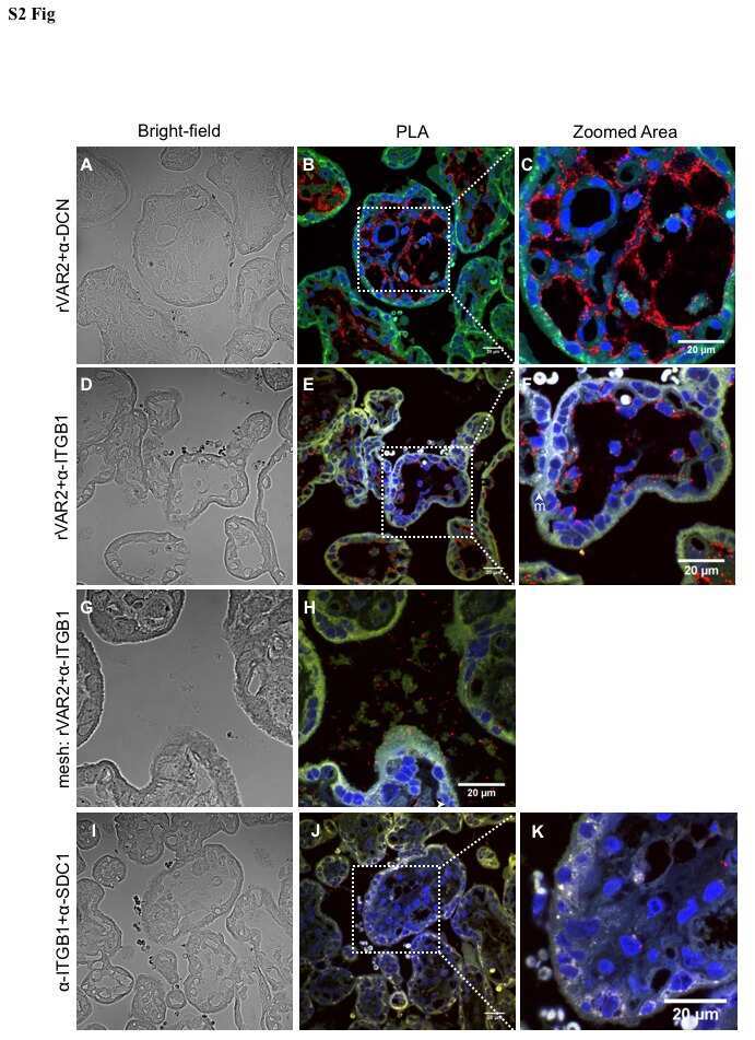
Placental Sequestration of Plasmodium falciparum Malaria Parasites Is Mediated by the Interaction Between VAR2CSA and Chondroitin Sulfate A on Syndecan-1.
Bang-Christensen SR, Pedersen RS, Pereira MA, Clausen TM, Løppke C, Sand NT, Ahrens TD, Jørgensen AM, Lim YC, Goksøyr L, Choudhary S, Gustavsson T, Dagil R, Daugaard M, Sander AF, Torp MH, Søgaard M, Theander TG, Østrup O, Lassen U, Hamerlik P, Salanti A, Agerbæk MØ
Cells 2019 Aug 28;8(9)
Cells 2019 Aug 28;8(9)
Leptospira interrogans causes quantitative and morphological disturbances in adherens junctions and other biological groups of proteins in human endothelial cells.
Sato H, Coburn J
PLoS neglected tropical diseases 2017 Jul;11(7):e0005830
PLoS neglected tropical diseases 2017 Jul;11(7):e0005830
Placental Sequestration of Plasmodium falciparum Malaria Parasites Is Mediated by the Interaction Between VAR2CSA and Chondroitin Sulfate A on Syndecan-1.
Ayres Pereira M, Mandel Clausen T, Pehrson C, Mao Y, Resende M, Daugaard M, Riis Kristensen A, Spliid C, Mathiesen L, E Knudsen L, Damm P, G Theander T, R Hansson S, A Nielsen M, Salanti A
PLoS pathogens 2016 Aug;12(8):e1005831
PLoS pathogens 2016 Aug;12(8):e1005831
No comments: Submit comment
Supportive validation
- Submitted by
- Invitrogen Antibodies (provider)
- Main image
- Experimental details
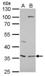
- Decorin Polyclonal Antibody detects DCN protein by western blot analysis. A. 30 µg A549 whole cell lysate/extract. B. 30 µg H1299 whole cell lysate/extract.10% SDS-PAGE. Decorin Polyclonal Antibody (Product # PA5-27370) dilution: 1:1,000. The HRP-conjugated anti-rabbit IgG antibody was used to detect the primary antibody.

- Submitted by
- Invitrogen Antibodies (provider)
- Main image
- Experimental details
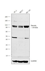
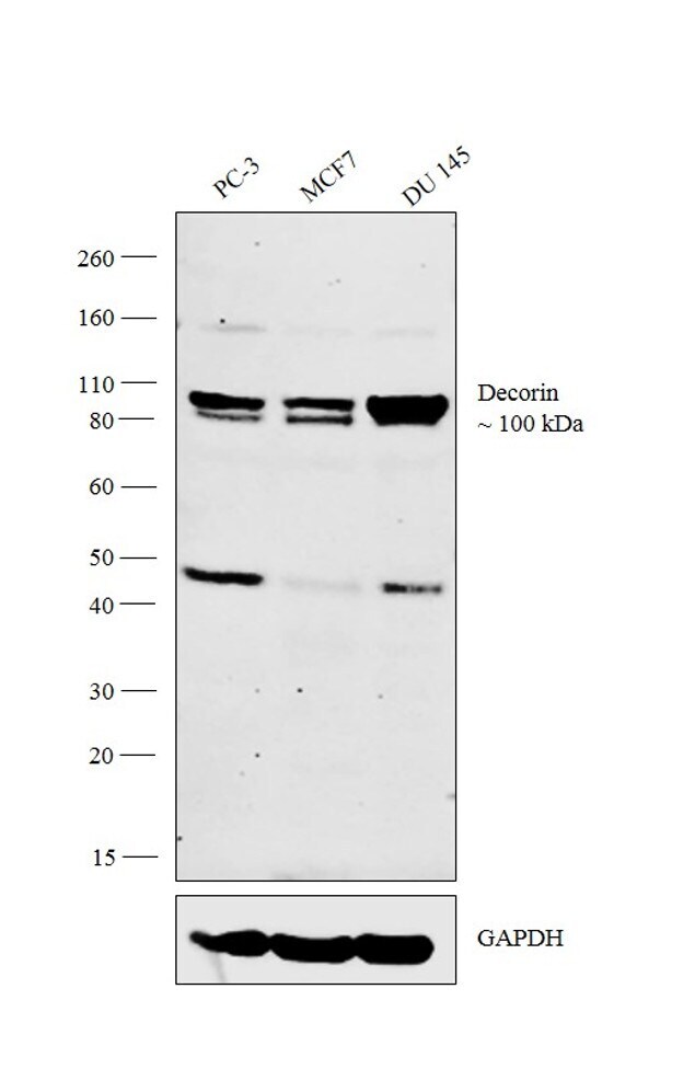
- Western blot analysis was performed on whole cell extracts (30 µg lysate) of PC-3 (Lane 1), MCF7 (Lane 2) and DU 145 (Lane 3). The blot was probed with Anti-Decorin Polyclonal Antibody (Product # PA5-27370, 1:1,000 dilution) and detected by chemiluminescence using Goat anti-Rabbit IgG (Heavy Chain) Superclonal™ Secondary Antibody, HRP conjugate (Product # A27036, 0.25 µg/mL, 1:4,000 dilution). A 100 kDa band corresponding to Decorin was observed across the cell lines tested.
Supportive validation
- Submitted by
- Invitrogen Antibodies (provider)
- Main image
- Experimental details
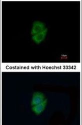
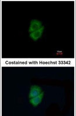
- Immunofluorescent analysis of Decorin in methanol-fixed A549 cells using a Decorin polyclonal antibody (Product # PA5-27370) at a 1:200 dilution.
Supportive validation
- Submitted by
- Invitrogen Antibodies (provider)
- Main image
- Experimental details
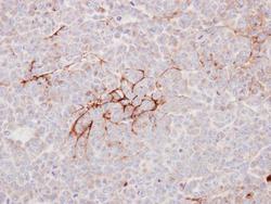
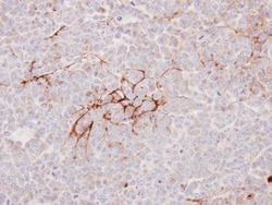
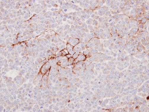
- Immunohistochemical analysis of paraffin-embedded TOV112D xenograft, using Decorin (Product # PA5-27370) antibody at 1:100 dilution. Antigen Retrieval: EDTA based buffer, pH 8.0, 15 min.
- Submitted by
- Invitrogen Antibodies (provider)
- Main image
- Experimental details
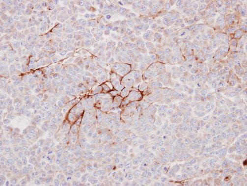
- Immunohistochemical analysis of paraffin-embedded TOV112D xenograft, using Decorin (Product # PA5-27370) antibody at 1:100 dilution. Antigen Retrieval: EDTA based buffer, pH 8.0, 15 min.
Supportive validation
- Submitted by
- Invitrogen Antibodies (provider)
- Main image
- Experimental details
- NULL
- Submitted by
- Invitrogen Antibodies (provider)
- Main image
- Experimental details
- NULL
- Submitted by
- Invitrogen Antibodies (provider)
- Main image
- Experimental details
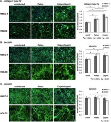
- Fig 1 Effect of Leptospira infection on extracellular matrix proteins in endothelial cells detected by immunofluorescence microscopy. (A) collagen type IV, (B) decorin, and (C) laminin in HMEC-1 and HDLEC are shown in green. The nuclei are stained in blue for all panels. Scale bars represent 50 mum. Quantified signal intensity of the host protein is indicated in the right-hand graphs (mean +/- SD, p -value is indicated below each graph, the independent p -values shown as an asterisk are compared to uninfected cells).