 Explore
 Validate
 Learn
Learn Western blot
Antibody data
- Antibody Data
- Antigen structure
- References [2]
- Comments [0]
- Validations
- Western blot [3]
- Immunocytochemistry [2]
Submit
Validation data
Reference
Comment
Report error
- Product number
- MAB8028 - Provider product page

- Provider
- R&D Systems
- Product name
- Human/Mouse/Rat p62/SQSTM1 Antibody
- Antibody type
- Monoclonal
- Description
- Protein A or G purified from hybridoma culture supernatant. Detects human p62/SQSTM1 in ELISAs. Detects human, mouse and rat p62/SQSTM1 in Western blots
- Reactivity
- Human, Mouse, Rat
- Host
- Mouse
- Conjugate
- Unconjugated
- Antigen sequence
Q13501- Isotype
- IgG
- Antibody clone number
- 864807
- Vial size
- 100 ug
- Storage
- Use a manual defrost freezer and avoid repeated freeze-thaw cycles. 12 months from date of receipt, -20 to -70 °C as supplied. 1 month, 2 to 8 °C under sterile conditions after reconstitution. 6 months, -20 to -70 °C under sterile conditions after reconstitution.
Submitted references Cold-inducible RNA-binding protein through TLR4 signaling induces mitochondrial DNA fragmentation and regulates macrophage cell death after trauma.
Tribbles Pseudokinase 3 Induces Both Apoptosis and Autophagy in Amyloid-β-induced Neuronal Death.
Li Z, Fan EK, Liu J, Scott MJ, Li Y, Li S, Xie W, Billiar TR, Wilson MA, Jiang Y, Wang P, Fan J
Cell death & disease 2017 May 11;8(5):e2775
Cell death & disease 2017 May 11;8(5):e2775
Tribbles Pseudokinase 3 Induces Both Apoptosis and Autophagy in Amyloid-β-induced Neuronal Death.
Saleem S, Biswas SC
The Journal of biological chemistry 2017 Feb 17;292(7):2571-2585
The Journal of biological chemistry 2017 Feb 17;292(7):2571-2585
No comments: Submit comment
Supportive validation
- Submitted by
- R&D Systems (provider)
- Main image
- Experimental details
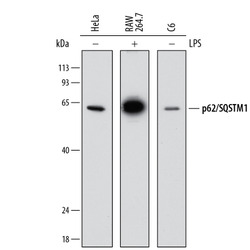
- Detection of Human, Mouse, and Rat p62/SQSTM1 by Western Blot. Western blot shows lysates of HeLa human cervical epithelial carcinoma cell line, RAW 264.7 mouse monocyte/macrophage cell line, and C6 rat glioma cell line untreated (-) or treated (+) with 1 µg/mL LPS for 24 hours. PVDF membrane was probed with 2 µg/mL of Mouse Anti-Human/Mouse/Rat p62/SQSTM1 Monoclonal Antibody (Catalog # MAB8028) followed by HRP-conjugated Anti-Mouse IgG Secondary Antibody (Catalog # HAF018). A specific band was detected for p62/SQSTM1 at approximately 62 kDa (as indicated). This experiment was conducted under reducing conditions and using Immunoblot Buffer Group 1.
- Submitted by
- R&D Systems (provider)
- Main image
- Experimental details
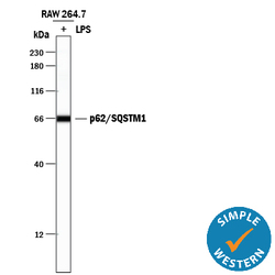
- Detection of Mouse p62/SQSTM1 by Simple WesternTM. Simple Western lane view shows lysates of RAW 264.7 mouse monocyte/macrophage cell line untreated (-) or treated (+) with 1 µg/mL LPS for 24 hours, loaded at 0.2 mg/mL. A specific band was detected for p62/SQSTM1 at approximately 66 kDa (as indicated) using 20 µg/mL of Mouse Anti-Human/Mouse/Rat p62/SQSTM1 Monoclonal Antibody (Catalog # MAB8028). This experiment was conducted under reducing conditions and using the 12-230 kDa separation system.

- Submitted by
- R&D Systems (provider)
- Main image
- Experimental details
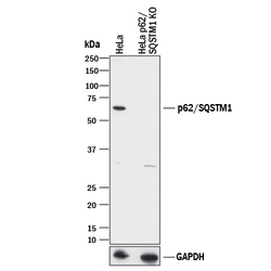
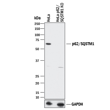
- Western Blot Shows Human p62/SQSTM1 Specificity Using Knockout Cell Line. Western blot shows lysates of HeLa human cervical epithelial carcinoma parental cell line and p62/SQSTM1 knockout HeLa cell line (KO). PVDF membrane was probed with 2 µg/mL of Mouse Anti-Human/Mouse/Rat p62/SQSTM1 Monoclonal Antibody (Catalog # MAB8028) followed by HRP-conjugated Anti-Mouse IgG Secondary Antibody (Catalog # HAF018). A specific band was detected for p62/SQSTM1 at approximately 62 kDa (as indicated) in the parental HeLa cell line, but is not detectable in the knockout HeLa cell line. GAPDH (Catalog # MAB5718) is shown as a loading control. This experiment was conducted under reducing conditions and using Immunoblot Buffer Group 1.
Supportive validation
- Submitted by
- R&D Systems (provider)
- Main image

- Experimental details
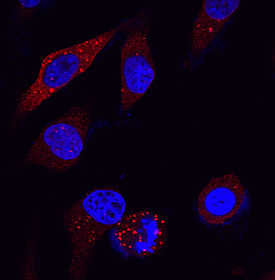
- p62/SQSTM1 in HeLa Human Cell Line. p62/SQSTM1 was detected in immersion fixed HeLa human cervical epithelial carcinoma cell line using Mouse Anti-Human/Mouse/Rat p62/SQSTM1 Monoclonal Antibody (Catalog # MAB8028) at 25 µg/mL for 3 hours at room temperature. Cells were stained using the NorthernLights™ 557-conjugated Anti-Mouse IgG Secondary Antibody (red; Catalog # NL007) and counterstained with DAPI (blue). Specific staining was localized to phagosomes in cell cytoplasm. View our protocol for Fluorescent ICC Staining of Cells on Coverslips.
- Submitted by
- R&D Systems (provider)
- Main image
- Experimental details
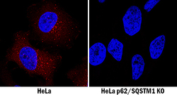
- p62/SQSTM1 Specificity is Shown by Immunocytochemistry in Knockout Cell Line. p62/SQSTM1 was detected in immersion fixed HeLa human cervical epithelial carcinoma cell line but is not detected in p62/SQSTM1 knockout (KO) HeLa cell line using Mouse Anti-Human/Mouse/Rat p62/SQSTM1 Monoclonal Antibody (Catalog # MAB8028) at 3 µg/mL for 3 hours at room temperature. Cells were stained using the NorthernLights 557-conjugated Anti-Mouse IgG Secondary Antibody (red; Catalog # NL007) and counterstained with DAPI (blue). Specific staining was localized to cytoplasm. View our protocol for Fluorescent ICC Staining of Cells on Coverslips.
