 Explore
 Validate
 Learn
Learn Western blot
Antibody data
- Antibody Data
- Antigen structure
- References [0]
- Comments [0]
- Validations
- Western blot [2]
- Immunocytochemistry [2]
Submit
Validation data
Reference
Comment
Report error
- Product number
- 710539 - Provider product page

- Provider
- Invitrogen Antibodies
- Product name
- SQSTM1 Recombinant Polyclonal Antibody (11HCLC)
- Antibody type
- Polyclonal
- Antigen
- Synthetic peptide
- Reactivity
- Human
- Host
- Rabbit
- Isotype
- IgG
- Antibody clone number
- 11HCLC
- Vial size
- 100 µg
- Concentration
- 0.5 mg/mL
- Storage
- Store at 4°C short term. For long term storage, store at -20°C, avoiding freeze/thaw cycles.
No comments: Submit comment
Supportive validation
- Submitted by
- Invitrogen Antibodies (provider)
- Main image
- Experimental details
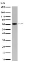
- Western blot analysis of Sequestosome-1 in whole extracts of U2OS treated with Chloroquinone at 60uM overnight using a Sequestosome-1 Recombinant Rabbit Polyclonal Antibody (Product # 710539) at a dilution of 1 µg/mL. Detection was performed using an HRP-conjugated Goat anti-Rabbit secondary antibody followed by chemiluminescence (ECL). Results show a band at ~62kDa.

- Submitted by
- Invitrogen Antibodies (provider)
- Main image
- Experimental details
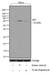
- Western blot analysis of p62 was performed by loading 20 µg of HeLa (lane1), serum starved HeLa (lane2), HeLa treated overnight with 10 mM of Rapamycin (lane3) cell lysate using Novex®NuPAGE® 12% Bis-Tris gel (Product # NP0342BOX), XCell SureLock Electrophoresis System (Product # EI0002), Novex® Sharp Pre-Stained Protein Standard (Product # LC5800), and iBlot® Dry Blotting System (Product # IB21001). Proteins were transferred to a nitrocellulose membrane and blocked with 5% skim milk for 1 hour at room temperature. p62 was detected at ~ 60 kDa using p62 Recombinant Rabbit Polyclonal Antibody (Product # 710539) at 1-3 µg/mL in 2.5% skim milk at 4°C overnight on a rocking platform. Goat anti-Rabbit IgG - HRP Secondary Antibody (Product # G-21234) at 1:5000 dilution was used and chemiluminescent detection was performed using Pierce™ ECL Western blotting Substrate (Product # 32106).

Supportive validation
- Submitted by
- Invitrogen Antibodies (provider)
- Main image
- Experimental details
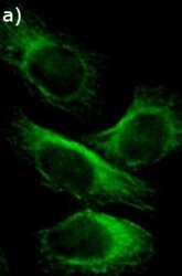
- Immunofluorescent analysis of Sequestosome-1 in HeLa cells using a Sequestosome-1 Recombinant Rabbit Polyclonal Antibody (Product # 710539) followed by detection using an Alexa Fluor 488-conjugated Goat anti-Rabbit secondary antibody (green).

- Submitted by
- Invitrogen Antibodies (provider)
- Main image

- Experimental details
- Immunofluorescent analysis of p62 was done on 70% confluent log phase HeLa cells. The cells were fixed with 4% paraformaldehyde for 15 minutes, permeabilized with 0.25% Triton X-100 for 10 minutes, and blocked with 5% BSA for 1 hour at room temperature. The cells were labeled with p62 Recombinant Rabbit Polyclonal Antibody (Product # 710539) at 2 µg/mL in 1% BSA and incubated for 3 hours at room temperature and then labeled with Alexa Fluor 488 Goat anti-Rabbit IgG Secondary Antibody (Product # A-11008) at a dilution of 1:400 for 30 minutes at room temperature (Panel a: green). Nuclei (Panel b: blue) were stained with SlowFade® Gold Antifade Mountant with DAPI (Product # S36938). F-actin (Panel c: red) was stained with Alexa Fluor 594 Phalloidin (Product # A12381). Panel d is a merged image showing cytoplasmic localization. Panel e shows no primary antibody control. The images were captured at 20X magnification.
