Explore
 Validate
 Learn
Learn Western blot
Antibody data
- Antibody Data
- Antigen structure
- References [1]
- Comments [0]
- Validations
- Western blot [5]
- Immunocytochemistry [6]
- Immunohistochemistry [2]
- Flow cytometry [4]
- Other assay [1]
Submit
Validation data
Reference
Comment
Report error
- Product number
- MA5-27800 - Provider product page

- Provider
- Invitrogen Antibodies
- Product name
- SQSTM1 Monoclonal Antibody (GT1478)
- Antibody type
- Monoclonal
- Antigen
- Recombinant full-length protein
- Description
- Positive Control: HepG2, A549, H1299, HCT116, Huh-7 (untreated), Huh-7 (3 µM Thapsigargin treatment for 12 hr), Huh-7 (3 µM Thapsigargin treatment for 24 hr), HepG2 (3 µM Thapsigargin treatment for 12 hr), HepG2 (3 µM Thapsigargin treatment for 24 hr), 2C4, MEF, MNT-1 Store product as a concentrated solution. Centrifuge briefly prior to opening the vial.
- Reactivity
- Human, Mouse
- Host
- Mouse
- Isotype
- IgG
- Antibody clone number
- GT1478
- Vial size
- 100 μL
- Concentration
- 1 mg/mL
- Storage
- Store at 4°C short term. For long term storage, store at -20°C, avoiding freeze/thaw cycles.
Submitted references Influences of cyclosporin A and non-immunosuppressive derivatives on cellular cyclophilins and viral nucleocapsid protein during human coronavirus 229E replication.
Ma-Lauer Y, Zheng Y, Malešević M, von Brunn B, Fischer G, von Brunn A
Antiviral research 2020 Jan;173:104620
Antiviral research 2020 Jan;173:104620
No comments: Submit comment
Supportive validation
- Submitted by
- Invitrogen Antibodies (provider)
- Main image
- Experimental details
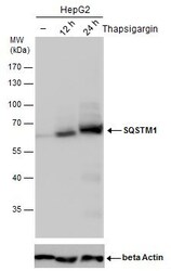
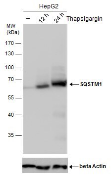
- SQSTM1 antibody detects SQSTM1 protein by western blot analysis. Un-treated (-) and treated (+, Thapsigargin treatment for 12 hr and 24hr) HepG2 whole cell extracts (30 µg) were separated by 10% SDS-PAGE, and the membrane was blotted with SQSTM1 antibody SQSTM1 Monoclonal Antibody (GT1478) (Product # MA5-27800) diluted by 1:1,000. The ACTB was used as internal control (Product # MA1-20221)4, 1:50,000) shown at the bottom panel. The HRP-conjugated anti-mouse IgG antibody was used to detect the primary antibody.
- Submitted by
- Invitrogen Antibodies (provider)
- Main image
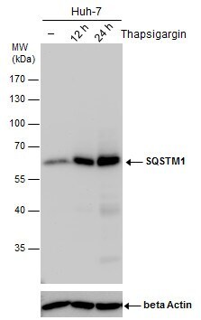
- Experimental details
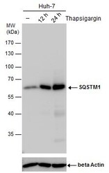
- SQSTM1 antibody detects SQSTM1 protein by western blot analysis. Un-treated (-) and treated (+, Thapsigargin treatment for 12 hr and 24hr) Huh-7 whole cell extracts (30 µg) were separated by 10% SDS-PAGE, and the membrane was blotted with SQSTM1 antibody SQSTM1 Monoclonal Antibody (GT1478) (Product # MA5-27800) diluted by 1:1,000. The ACTB was used as internal control (Product # MA1-20221)4, 1:50,000) shown at the bottom panel. The HRP-conjugated anti-mouse IgG antibody was used to detect the primary antibody.
- Submitted by
- Invitrogen Antibodies (provider)
- Main image
- Experimental details
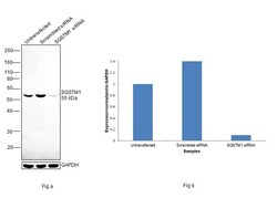
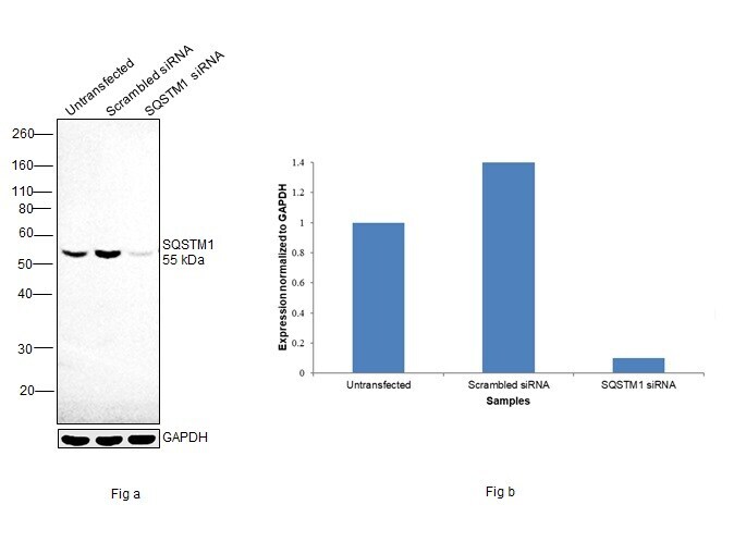
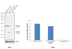
- Knockdown of SQSTM1 was achieved by transfecting Hep G2 with SQSTM1 specific siRNAs (Silencer® select Product # S16960, S16962). Western blot analysis (Fig. a) was performed using Whole cell extracts from the SQSTM1 knockdown cells (lane 3), non-targeting scrambled siRNA transfected cells (lane 2) and untransfected cells (lane 1). The blot was probed with SQSTM1 Monoclonal Antibody (GT1478) (Product # MA5-27800, 1:1000 dilution) and Goat anti-Mouse IgG (H+L) Superclonal™ Recombinant Secondary Antibody, HRP (Product # A28177, 1:4000 dilution). Densitometric analysis of this western blot is shown in histogram (Fig. b). Decrease in signal upon siRNA mediated knock down confirms that antibody is specific to SQSTM1.
- Submitted by
- Invitrogen Antibodies (provider)
- Main image
- Experimental details
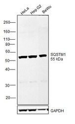
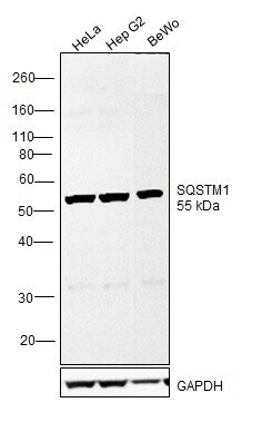
- Western blot was performed using Anti-SQSTM1 Monoclonal Antibody (GT1478) (Product # MA5-27800) and a ~55 kDa band corresponding to SQSTM1 was observed across cell lines tested. Whole cell extracts (30 µg lysate) of HeLa (Lane 1), Hep G2 (Lane 2) and BeWo (Lane 3) were electrophoresed using NuPAGE™ 10% Bis-Tris Protein Gel (Product # NP0301BOX). Resolved proteins were then transferred onto a Nitrocellulose membrane (Product # IB23001) by iBlot® 2 Dry Blotting System (Product # IB21001). The blot was probed with the primary antibody (1:1000 dilution) and detected by chemiluminescence with Goat anti-Mouse IgG (H+L) Superclonal™ Recombinant Secondary Antibody, HRP (Product # A28177,1:4000 dilution) using the iBright FL 1000 (Product # A32752). Chemiluminescent detection was performed using Novex® ECL Chemiluminescent Substrate Reagent Kit (Product # WP20005).
- Submitted by
- Invitrogen Antibodies (provider)
- Main image
- Experimental details
- Knockout of SQSTM1 was achieved by CRISPR-Cas9 genome editing using LentiArray™ Lentiviral sgRNA (Product # A32042, Assay ID CRISPR970861_LV) and LentiArray Cas9 Lentivirus (Product # A32064). Western blot analysis of SQSTM1 was performed by loading 30 µg of HeLa Wild Type (Lane 1), HeLa Cas9 (Lane 2) andHela SQSTM1 KO (Lane 3) whole cell extracts. The samples were electrophoresed using NuPAGE™ Novex™ 4-12% Bis-Tris Protein Gel (Product # NP0322BOX). Resolved proteins were then transferred onto a nitrocellulose membrane (Product # IB23001) by iBlot® 2 Dry Blotting System (Product # IB21001). The blot was probed with Anti-SQSTM1 Monoclonal Antibody (GT1478) (Product # MA5-27800, 1:1,000 dilution) and Goat anti-Mouse IgG (H+L) Superclonal™ Recombinant Secondary Antibody, HRP (Product # A28177, 1:6,000 dilution) using the iBright FL 1000 (Product # A32752). Chemiluminescent detection was performed using Novex® ECL Chemiluminescent Substrate Reagent Kit (Product # WP20005). Loss of signal upon CRISPR mediated knockout (KO) using the LentiArray™ CRISPR product line confirms that antibody is specific to SQSTM1.

Supportive validation
- Submitted by
- Invitrogen Antibodies (provider)
- Main image
- Experimental details
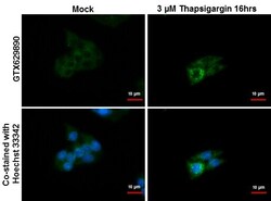
- Immunocytochemistry-Immunofluorescence analysis of SQSTM1 was performed in HepG2 cells treated with 3 µM thapsigargin 16 hrs (rigtht) and mock (left) fixed in ice-cold MeOH for 10 Min, permeabilize with cooled acetone for 1 min . Green: SQSTM1 Monoclonal Antibody (GT1478) (Product # MA5-27800) diluted at 1:500. Blue: Hoechst 33342 staining.

- Submitted by
- Invitrogen Antibodies (provider)
- Main image

- Experimental details
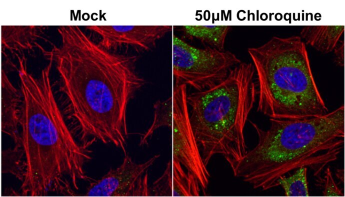
- SQSTM1 antibody [GT1478] detects SQSTM1 protein at autophagosome by immunofluorescent analysis. Samples: HeLa cells mock (left) and treated with 50 µM Chloroquine for 24 hr (right) were fixed in 4% paraformaldehyde at RT for 15 min. Green: SQSTM1 protein stained by SQSTM1 antibody [GT1478] (Product # MA5-27800) diluted at 1:1,000. Red: phalloidin, a cytoskeleton marker, stained by phalloidin (invitrogen, A12380) diluted at 1:200. Blue: Hoechst 33342 staining.

- Submitted by
- Invitrogen Antibodies (provider)
- Main image

- Experimental details
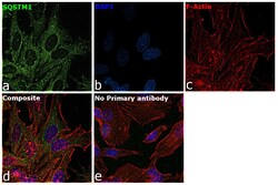
- Immunofluorescence analysis of SQSTM1 was performed using 70% confluent log phase HeLa cells. The cells were fixed with 4% paraformaldehyde for 10 minutes, permeabilized with 0.1% Triton™ X-100 for 15 minutes, and blocked with 2% BSA for 45 minutes at room temperature. The cells were labeled with SQSTM1 Monoclonal Antibody (GT1478) (Product # MA5-27800) at 1:500 dilution in 0.1% BSA, incubated at 4 degree celsius overnight and then labeled with Goat anti-Mouse IgG (H+L) Highly Cross-Adsorbed Secondary Antibody, Alexa Fluor Plus 488 (Product # A32723), (1:2000 dilution), for 45 minutes at room temperature (Panel a: Green). Nuclei (Panel b:Blue) were stained with ProLong™ Diamond Antifade Mountant with DAPI (Product # P36962). F-actin (Panel c: Red) was stained with Rhodamine Phalloidin (Product # R415, 1:300 dilution). Panel d represents the merged image showing cytoplasm, endosome-like, plasma membrane and nuclear localization. Panel e represents control cells with no primary antibody to assess background. The images were captured at 60X magnification.

- Submitted by
- Invitrogen Antibodies (provider)
- Main image

- Experimental details
- SQSTM1 antibody [GT1478] detects SQSTM1 protein at autophagosome by immunofluorescent analysis. Samples: HeLa cells mock (left) and treated with 50 µM Chloroquine for 24 hr (right) were fixed in 4% paraformaldehyde at RT for 15 min. Green: SQSTM1 protein stained by SQSTM1 antibody [GT1478] (Product # MA5-27800) diluted at 1:1,000. Red: phalloidin, a cytoskeleton marker, stained by phalloidin (invitrogen, A12380) diluted at 1:200. Blue: Hoechst 33342 staining.
- Submitted by
- Invitrogen Antibodies (provider)
- Main image

- Experimental details
- Immunocytochemistry-Immunofluorescence analysis of SQSTM1 was performed in HepG2 cells treated with 3 µM thapsigargin 16 hrs (rigtht) and mock (left) fixed in ice-cold MeOH for 10 Min, permeabilize with cooled acetone for 1 min . Green: SQSTM1 Monoclonal Antibody (GT1478) (Product # MA5-27800) diluted at 1:500. Blue: Hoechst 33342 staining.

- Submitted by
- Invitrogen Antibodies (provider)
- Main image
- Experimental details
- Immunofluorescence analysis of SQSTM1 was performed using 70% confluent log phase HeLa cells. The cells were fixed with 4% paraformaldehyde for 10 minutes, permeabilized with 0.1% Triton™ X-100 for 15 minutes, and blocked with 2% BSA for 45 minutes at room temperature. The cells were labeled with SQSTM1 Monoclonal Antibody (GT1478) (Product # MA5-27800) at 1:500 dilution in 0.1% BSA, incubated at 4 degree celsius overnight and then labeled with Goat anti-Mouse IgG (H+L) Highly Cross-Adsorbed Secondary Antibody, Alexa Fluor Plus 488 (Product # A32723), (1:2000 dilution), for 45 minutes at room temperature (Panel a: Green). Nuclei (Panel b:Blue) were stained with ProLong™ Diamond Antifade Mountant with DAPI (Product # P36962). F-actin (Panel c: Red) was stained with Rhodamine Phalloidin (Product # R415, 1:300 dilution). Panel d represents the merged image showing cytoplasm, endosome-like, plasma membrane and nuclear localization. Panel e represents control cells with no primary antibody to assess background. The images were captured at 60X magnification.
Supportive validation
- Submitted by
- Invitrogen Antibodies (provider)
- Main image

- Experimental details
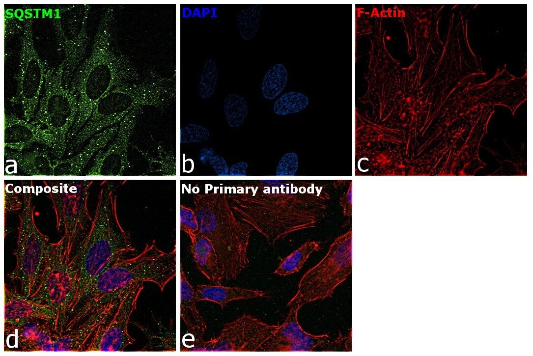
- SQSTM1 antibody [GT1478] detects SQSTM1 protein expression by immunohistochemical analysis. Sample: Frozen sectioned adult mouse retina. Green: SQSTM1 protein stained by SQSTM1 antibody [GT1478] (Product # MA5-27800) diluted at 1:250. Red: beta Tubulin 3/ TUJ1, stained by beta Tubulin 3/ TUJ1 antibody [GT11710] diluted at 1:250. Blue: Fluoroshield with DAPI .

- Submitted by
- Invitrogen Antibodies (provider)
- Main image
- Experimental details
- SQSTM1 antibody [GT1478] detects SQSTM1 protein expression by immunohistochemical analysis. Sample: Frozen sectioned adult mouse retina. Green: SQSTM1 protein stained by SQSTM1 antibody [GT1478] (Product # MA5-27800) diluted at 1:250. Red: beta Tubulin 3/ TUJ1, stained by beta Tubulin 3/ TUJ1 antibody [GT11710] diluted at 1:250. Blue: Fluoroshield with DAPI .

Supportive validation
- Submitted by
- Invitrogen Antibodies (provider)
- Main image

- Experimental details
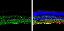
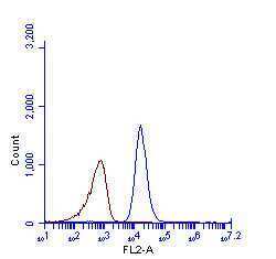
- Flow cytometry analysis of SQSTM1 in HeLa cells. Samples were treated with SQSTM1 monoclonal antibody (Product # MA5-27800) using a dilution of 1:100. Results are as follows: Brown: Unlabelled sample was also used as a control, Blue: SQSTM1 antibody. Acquisition of >20,000 events were collected using Argon ion laser (488nm) and 525/30 bandpass filter..
- Submitted by
- Invitrogen Antibodies (provider)
- Main image
- Experimental details
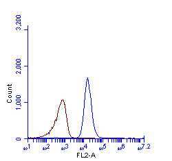
- SQSTM1 antibody [GT1478] (Product # MA5-27800) detects SQSTM1 protein by flow cytometry analysis. Sample: HeLa cell fixed in 4% paraformaldehyde at 4°C for 5 min. Brown: Unlabelled sample was also used as a control. Blue: SQSTM1 antibody [GT1478] (Product # MA5-27800) dilution: 1:100. Acquisition of >20,000 events were collected using Argon ion laser (488nm) and 525/30 bandpass filter.
- Submitted by
- Invitrogen Antibodies (provider)
- Main image

- Experimental details
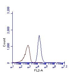
- SQSTM1 antibody [GT1478] (Product # MA5-27800) detects SQSTM1 protein by flow cytometry analysis. Sample: HeLa cell fixed in 4% paraformaldehyde at 4°C for 5 min. Brown: Unlabelled sample was also used as a control. Blue: SQSTM1 antibody [GT1478] (Product # MA5-27800) dilution: 1:100. Acquisition of >20,000 events were collected using Argon ion laser (488nm) and 525/30 bandpass filter.
- Submitted by
- Invitrogen Antibodies (provider)
- Main image
- Experimental details
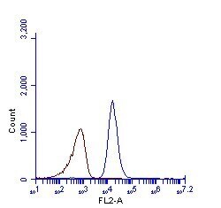
- SQSTM1 antibody [GT1478] (Product # MA5-27800) detects SQSTM1 protein by flow cytometry analysis. Sample: HeLa cell fixed in 4% paraformaldehyde at 4°C for 5 min. Brown: Unlabelled sample was also used as a control. Blue: SQSTM1 antibody [GT1478] (Product # MA5-27800) dilution: 1:100. Acquisition of >20,000 events were collected using Argon ion laser (488nm) and 525/30 bandpass filter.

Supportive validation
- Submitted by
- Invitrogen Antibodies (provider)
- Main image
- Experimental details
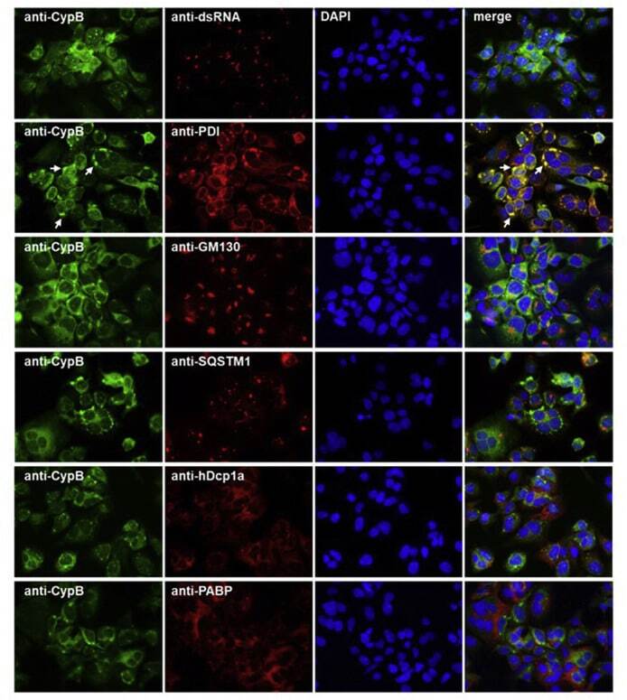
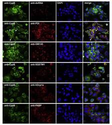
- Fig. 6 Co-immunostaining of CypB and cell organelles in HCoV-229E-infected Huh7 cells . For the identification of the intense cyclophilin B bleb-like structures infected cells (MOI = 1; 48 h p.i.) were co-stained with anti-CypB and antibodies directed against markers of the ER (anti-PDI), cis-GOLGI (anti-GM130), autophagosomes (anti-SQSTM1), anti-P-bodies (anti-hDcp1a) and stress granules (anti-PABP). Cyclophilin B normally distributes within the ER. In the presence of HCoV-229E, it intriguingly concentrates at bleb-like structures of the ER. Fig. 6