 Explore
 Validate
 Learn
Learn Western blot
 Immunocytochemistry
Antibody data
- Antibody Data
- Antigen structure
- References [3]
- Comments [0]
- Validations
- Western blot [1]
Submit
Validation data
Reference
Comment
Report error
- Product number
- NBP1-42821 - Provider product page

- Provider
- Novus Biologicals
- Proper citation
- Novus Cat#NBP1-42821, RRID:AB_10005939
- Product name
- Rabbit Polyclonal p62/SQSTM1 Antibody
- Antibody type
- Polyclonal
- Description
- Immunogen affinity purified.
- Reactivity
- Human, Mouse, Rat
- Host
- Rabbit
- Isotype
- IgG
- Vial size
- 0.1 ml
- Concentration
- 1 mg/ml
- Storage
- Store at 4C short term. Aliquot and store at -20C long term. Avoid freeze-thaw cycles.
Submitted references Modulating EGFR-MTORC1-autophagy as a potential therapy for persistent fetal vasculature (PFV) disease.
SMCR8 negatively regulates AKT and MTORC1 signaling to modulate lysosome biogenesis and tissue homeostasis.
The helminth product, ES-62 modulates dendritic cell responses by inducing the selective autophagolysosomal degradation of TLR-transducers, as exemplified by PKCδ.
Yazdankhah M, Shang P, Ghosh S, Bhutto IA, Stepicheva N, Grebe R, Hose S, Weiss J, Luo T, Mishra S, Riazuddin SA, Ghosh A, Handa JT, Lutty GA, Zigler JS Jr, Sinha D
Autophagy 2020 Jun;16(6):1130-1142
Autophagy 2020 Jun;16(6):1130-1142
SMCR8 negatively regulates AKT and MTORC1 signaling to modulate lysosome biogenesis and tissue homeostasis.
Lan Y, Sullivan PM, Hu F
Autophagy 2019 May;15(5):871-885
Autophagy 2019 May;15(5):871-885
The helminth product, ES-62 modulates dendritic cell responses by inducing the selective autophagolysosomal degradation of TLR-transducers, as exemplified by PKCδ.
Eason RJ, Bell KS, Marshall FA, Rodgers DT, Pineda MA, Steiger CN, Al-Riyami L, Harnett W, Harnett MM
Scientific reports 2016 Nov 21;6:37276
Scientific reports 2016 Nov 21;6:37276
No comments: Submit comment
Supportive validation
- Submitted by
- Novus Biologicals (provider)
- Main image
- Experimental details
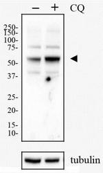
- Western Blot: p62/SQSTM1 Antibody [NBP1-42821] - HeLa cells were treated with (+) or without 50 uM (-) of Chloriquine (CQ) for 24 hours. Total cell lysates were prepared and separated on a 12% gel by SDS-PAGE. Protein was transferred to PVDF membrane and blocked in 5% non-fat milk. The membrane was then probed with 2 ug/ml anti-p62/SQSMT1 in 1% milk and detected with an anti-rabbit HRP secondary antibody using chemiluminescence. Note the upregulation of p62 (arrowhead) in response to chloroquine treatment and the blockage of autophagy.
