Explore
 Validate
 Learn
LearnPA5-27247
antibody from Invitrogen Antibodies
Targeting: SQSTM1
A170, OSIL, p60, p62, p62B, PDB3
 Western blot
 Immunocytochemistry Immunoprecipitation Immunohistochemistry Flow cytometry Other assay
Antibody data
- Antibody Data
- Antigen structure
- References [1]
- Comments [0]
- Validations
- Immunocytochemistry [2]
- Immunohistochemistry [1]
- Flow cytometry [2]
- Other assay [1]
Submit
Validation data
Reference
Comment
Report error
- Product number
- PA5-27247 - Provider product page

- Provider
- Invitrogen Antibodies
- Product name
- SQSTM1 Polyclonal Antibody
- Antibody type
- Polyclonal
- Antigen
- Synthetic peptide
- Description
- Recommended positive controls: A549, HepG2, HepG2 (3µM Thapsigargin treatment for 24 hr), Huh7, Huh7 ( 3µM Thapsigargin, 12hr), NIH-3T3, JC, BCL-1, PC-12, Rat2, HeLa. Store product as a concentrated solution. Centrifuge briefly prior to opening the vial.
- Reactivity
- Human, Mouse, Rat, Bovine
- Host
- Rabbit
- Isotype
- IgG
- Vial size
- 100 μL
- Concentration
- 0.39 mg/mL
- Storage
- Store at 4°C short term. For long term storage, store at -20°C, avoiding freeze/thaw cycles.
Submitted references Tau Oligomers and Fibrils Exhibit Differential Patterns of Seeding and Association With RNA Binding Proteins.
Jiang L, Zhao J, Cheng JX, Wolozin B
Frontiers in neurology 2020;11:579434
Frontiers in neurology 2020;11:579434
No comments: Submit comment
Supportive validation
- Submitted by
- Invitrogen Antibodies (provider)
- Main image

- Experimental details
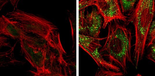
- SQSTM1 antibody [N3C1], Internal detects SQSTM1 protein at autophagosome by immunofluorescent analysis. Samples: HeLa cells mock (left) and treated with 50μM Chloroquine for 24 hr (right) were fixed in 4% paraformaldehyde at RT for 15 min. Green: SQSTM1 protein stained by SQSTM1 antibody [N3C1], Internal (Product # PA5-27247) diluted at 1:1,000. Red: Phalloidin, a F-actin marker.
- Submitted by
- Invitrogen Antibodies (provider)
- Main image

- Experimental details
- SQSTM1 antibody [N3C1], Internal detects SQSTM1 protein at autophagosome by immunofluorescent analysis. Samples: HeLa cells mock (left) and treated with 50μM Chloroquine for 24 hr (right) were fixed in 4% paraformaldehyde at RT for 15 min. Green: SQSTM1 protein stained by SQSTM1 antibody [N3C1], Internal (Product # PA5-27247) diluted at 1:1,000. Red: Phalloidin, a F-actin marker.

Supportive validation
- Submitted by
- Invitrogen Antibodies (provider)
- Main image

- Experimental details
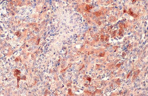
- SQSTM1/P62 antibody [N3C1], Internal detects SQSTM1/P62 protein at cytoplasm by immunohistochemical analysis. Sample: Paraffin-embedded human lung cancer. SQSTM1/P62 stained by SQSTM1/P62 antibody [N3C1], Internal (Product # PA5-27247) diluted at 1:500. Antigen Retrieval: Citrate buffer, pH 6.0, 15 min.
Supportive validation
- Submitted by
- Invitrogen Antibodies (provider)
- Main image

- Experimental details
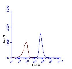
- SQSTM1 antibody [N3C1], Internal (Product # PA5-27247) detects SQSTM1 protein by flow cytometry analysis. Sample: HeLa cell fixed in 4% paraformaldehyde at 4°C for 5 min. Brown: Unlabelled sample was also used as a control. Blue: SQSTM1 antibody [N3C1], Internal] (Product # PA5-27247) dilution: 1:100. Acquisition of >20,000 events were collected using Argon ion laser (488nm) and 525/30 bandpass filter.
- Submitted by
- Invitrogen Antibodies (provider)
- Main image

- Experimental details
- SQSTM1 antibody [N3C1], Internal (Product # PA5-27247) detects SQSTM1 protein by flow cytometry analysis. Sample: HeLa cell fixed in 4% paraformaldehyde at 4°C for 5 min. Brown: Unlabelled sample was also used as a control. Blue: SQSTM1 antibody [N3C1], Internal] (Product # PA5-27247) dilution: 1:100. Acquisition of >20,000 events were collected using Argon ion laser (488nm) and 525/30 bandpass filter.

Supportive validation
- Submitted by
- Invitrogen Antibodies (provider)
- Main image

- Experimental details
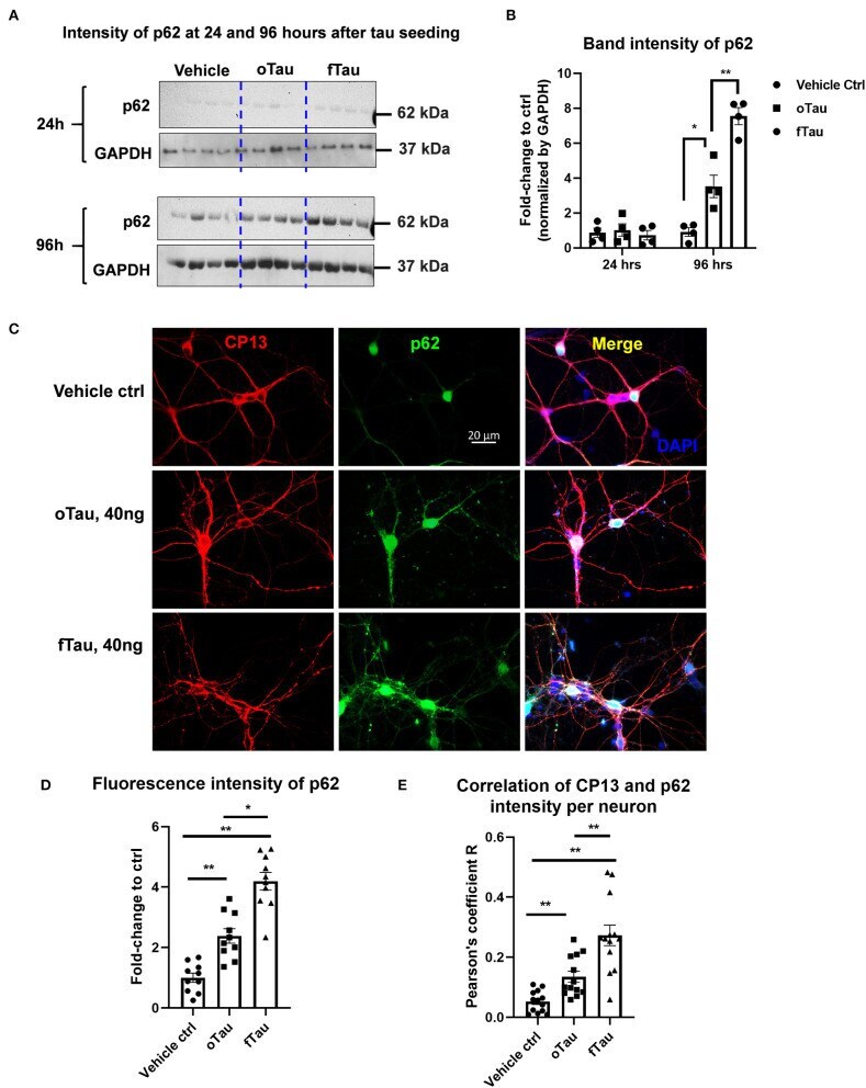
- Figure 5 Co-localization of large tau inclusions with p62. (A) Immunoblotting detecting p62 expression in neurons after oTau or fTau treatment. Each lane represents an independent biological replicate. The immunoblotting was performed with cell lysate from hippocampal cultures overexpressing human 4R0N WT tau and treated with vehicle, oTau, or fTau. The cells were harvested at 24 and 96 h after treatment. The p62 antibody was used to identify the expression level of p62 protein. (B) Quantification of p62 immunoblot in cell lysate of hippocampal cultures overexpressing human 4R0N WT tau, treated with vehicle control, oTau, or fTau on day 14, and harvested at 24 or 96 h after treatment. Data are shown as mean +- SEM, N = 4, data analysis was done by two-way ANOVA, multiple comparison test by Fisher's LSD, * p < 0.05, ** p < 0.01 in comparison with vehicle control. (C) Representative images showing the co-localization of phosphorylated tau inclusions CP13 (red) with p62 granules (green) at 96 h after oTau, fTau, or vehicle treatment in hippocampal neurons overexpressing human 4R0N tau. Co-labeling marker is DAPI (blue) to show the cell nucleus. Scale bar 20 mum. (D) Quantification of p62 fluorescence intensity in (C) , which is the immunofluorescence labeling of hippocampal cultures overexpressing human 4R0N WT tau, treated with vehicle control, oTau, or fTau on day 14, and harvested at 96 h after treatment. Data are shown as mean +- SEM, N = 10, data analysis was done by one-wa