 Explore
 Validate
 Learn
LearnMA5-14661
antibody from Invitrogen Antibodies
Targeting: SERPINA1
A1A, A1AT, AAT, alpha-1-antitrypsin, alpha1AT, PI, PI1
 Western blot
Antibody data
- Antibody Data
- Antigen structure
- References [0]
- Comments [0]
- Validations
- Western blot [4]
- Flow cytometry [1]
Submit
Validation data
Reference
Comment
Report error
- Product number
- MA5-14661 - Provider product page

- Provider
- Invitrogen Antibodies
- Product name
- alpha-1 Antitrypsin Monoclonal Antibody (TMF1#4B5)
- Antibody type
- Monoclonal
- Antigen
- Purifed from natural sources
- Description
- MA5-14661 targets Alpha1-Antitrypsin in ELISA, IP, and RIA applications and shows reactivity with Human samples.
- Antibody clone number
- TMF1#4B5
- Concentration
- 1 mg/mL
No comments: Submit comment
Supportive validation
- Submitted by
- Invitrogen Antibodies (provider)
- Main image
- Experimental details
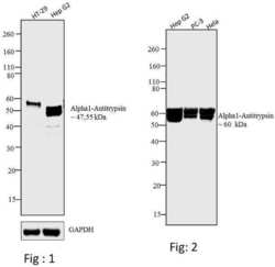
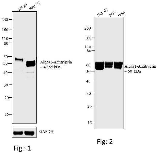
- Western blot analysis was performed on membrane enriched extracts (30 µg) of (Fig 1) HT-29 (Lane 1) and Hep G2 (Lane 2).Likewise, Western blot analysis was performed on (Fig 2) condition media of Hep G2 (Lane 1), PC3 (Lane 2) and HeLa (Lane 3).The blots were probed with Anti-Alpha1-Antitrypsin Mouse Monoclonal Antibody (Product # MA5-14661, 2 µg/mL) and detected by chemiluminescence using Goat anti-Mouse IgG (H+L) Superclonal™ Secondary Antibody, HRP conjugate (Product # A28177, 0.4 µg/mL, 1:2500 dilution). In Figure 1, a ~47 kDa band corresponding Alpha1-Antitrypsin was observed in Hep G2, and a ~55 kDa band was observed in HT-29 due to glycosylation of protein. Also, in Figure 2, ~60 kDa bands were observed in Hep G2, PC 3 and HeLa due to glycosylation of protein. Known quantity of protein samples were electrophoresed using Novex® NuPAGE®4-12 % Bis-Tris gel (Product # NP0321BOX), XCell SureLock™ Electrophoresis System (Product # EI0002) and Novex® Sharp Pre-Stained Protein Standard (Product # LC5800). Resolved proteins were then transferred onto a nitrocellulose membrane by iBlot® 2 Dry Blotting System (Product # IB21001). The membrane was probed with the relevant primary and secondary Antibody using iBind™ Flex Western Starter Kit (Product # SLF2000S). Chemiluminescent detection was performed using Pierce™ ECL Western Blotting Substrate (Product # 32106).
- Submitted by
- Invitrogen Antibodies (provider)
- Main image
- Experimental details
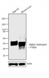
- Western blot analysis of Alpha1-Antitrypsin was performed using membrane enriched extracts (30 µg lysate) of Hep G2 (Lane 1) and tissue extract of Mouse Liver (Lane 2). The blots were probed with Anti-Alpha1-Antitrypsin Mouse Monoclonal Antibody (Product # MA5-14661, 2 µg/mL) and detected by chemiluminescence using Goat anti-Mouse IgG (H+L) Superclonal™ Secondary Antibody, HRP conjugate (Product # A28177, 0.4 µg/mL, 1:2500 dilution). A 47 kDa band corresponding to Alpha1-Antitrypsin was observed across the cell line and tissue tested. Known quantity of protein samples were electrophoresed using Novex® NuPAGE® 10 % Bis-Tris gel (Product # NP0302BOX), XCell SureLock™ Electrophoresis System (Product # EI0002) and Novex® Sharp Pre-Stained Protein Standard (Product # LC5800). Resolved proteins were then transferred onto a nitrocellulose membrane with iBlot® 2 Dry Blotting System (Product # IB21001). The membrane was probed with the relevant primary and secondary Antibody following blocking with 5 % skimmed milk. Chemiluminescent detection was performed using Pierce™ ECL Western Blotting Substrate (Product # 32106).

- Submitted by
- Invitrogen Antibodies (provider)
- Main image

- Experimental details
- Western blot analysis of Alpha1-Antitrypsin was performed using membrane enriched extracts (30 µg lysate) of Hep G2 (Lane 1) and tissue extract of Mouse Liver (Lane 2). The blots were probed with Anti-Alpha1-Antitrypsin Mouse Monoclonal Antibody (Product # MA5-14661, 2 µg/mL) and detected by chemiluminescence using Goat anti-Mouse IgG (H+L) Superclonal™ Secondary Antibody, HRP conjugate (Product # A28177, 0.4 µg/mL, 1:2500 dilution). A 47 kDa band corresponding to Alpha1-Antitrypsin was observed across the cell line and tissue tested. Known quantity of protein samples were electrophoresed using Novex® NuPAGE® 10 % Bis-Tris gel (Product # NP0302BOX), XCell SureLock™ Electrophoresis System (Product # EI0002) and Novex® Sharp Pre-Stained Protein Standard (Product # LC5800). Resolved proteins were then transferred onto a nitrocellulose membrane with iBlot® 2 Dry Blotting System (Product # IB21001). The membrane was probed with the relevant primary and secondary Antibody following blocking with 5 % skimmed milk. Chemiluminescent detection was performed using Pierce™ ECL Western Blotting Substrate (Product # 32106).

- Submitted by
- Invitrogen Antibodies (provider)
- Main image
- Experimental details
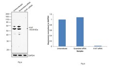
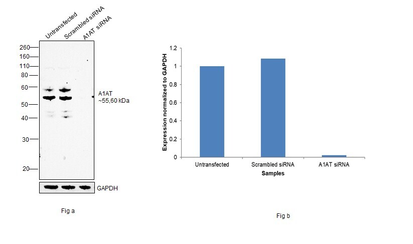
- Knockdown of alpha-1 Antitrypsin was achieved by transfecting Hep G2 with alpha-1 Antitrypsin specific siRNAs (Silencer® select Product # S10459, S10458). Western blot analysis (Fig. a) was performed using Membrane enriched extracts from the alpha-1 Antitrypsin knockdown cells (lane 3), non-targeting scrambled siRNA transfected cells (lane 2) and untransfected cells (lane 1). The blot was probed with alpha-1 Antitrypsin Monoclonal Antibody (TMF1#4B5) (Product # MA5-14661, 2µg/mL) and Goat anti-Mouse IgG (H+L) Superclonal™ Recombinant Secondary Antibody, HRP (Product # A28177, 1:4000 dilution). Densitometric analysis of this western blot is shown in histogram (Fig. b). Decrease in signal upon siRNA mediated knock down confirms that antibody is specific to alpha-1 Antitrypsin.
Supportive validation
- Submitted by
- Invitrogen Antibodies (provider)
- Main image
- Experimental details
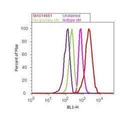
- Flow cytometry analysis of Alpha1-Antitrypsin was performed using Hep G2 cells. Cells were fixed with 70% ethanol for 10 minutes, permeabilized with 0.25% Triton™ X-100 for 20 minutes, and blocked with 5% BSA for 30 minutes at room temperature. Cells were labeled with Alpha1-Antitrypsin Mouse Monoclonal Antibody (Product # MA5-14661, red histogram) or with mouse isotype control (pink histogram) at 3-5 µg/million cells in 2.5% BSA. After incubation at room temperature for 2 hours, the cells were labeled with Alexa Fluor® 488 Rabbit Anti-Mouse Secondary Antibody (Product # A-11059) at a dilution of 1:400 for 30 minutes at room temperature. The representative 10,000 cells were acquired and analyzed for each sample using an Attune® Acoustic Focusing Cytometer. The purple histogram represents unstained control cells and the green histogram represents no-primary-antibody control.
