Explore
 Validate
 Learn
LearnMA5-15521
antibody from Invitrogen Antibodies
Targeting: SERPINA1
A1A, A1AT, AAT, alpha-1-antitrypsin, alpha1AT, PI, PI1
 Western blot
Antibody data
- Antibody Data
- Antigen structure
- References [2]
- Comments [0]
- Validations
- Western blot [5]
- Other assay [2]
Submit
Validation data
Reference
Comment
Report error
- Product number
- MA5-15521 - Provider product page

- Provider
- Invitrogen Antibodies
- Product name
- alpha-1 Antitrypsin Monoclonal Antibody (2B12)
- Antibody type
- Monoclonal
- Antigen
- Purifed from natural sources
- Description
- MA5-15521 targets AAT in WB applications and shows reactivity with Human and mouse samples.
- Antibody clone number
- 2B12
- Concentration
- Conc. Not Determined
Submitted references Pulmonary transplantation of alpha-1 antitrypsin (AAT)-transgenic macrophages provides a source of functional human AAT in vivo.
Improved bi-allelic modification of a transcriptionally silent locus in patient-derived iPSC by Cas9 nickase.
Janosz E, Hetzel M, Spielmann H, Tumpara S, Rossdam C, Schwabbauer M, Kloos D, von Kaisenberg C, Schambach A, Buettner FFR, Janciauskiene S, Lachmann N, Moritz T
Gene therapy 2021 Sep;28(9):477-493
Gene therapy 2021 Sep;28(9):477-493
Improved bi-allelic modification of a transcriptionally silent locus in patient-derived iPSC by Cas9 nickase.
Eggenschwiler R, Moslem M, Fráguas MS, Galla M, Papp O, Naujock M, Fonfara I, Gensch I, Wähner A, Beh-Pajooh A, Mussolino C, Tauscher M, Steinemann D, Wegner F, Petri S, Schambach A, Charpentier E, Cathomen T, Cantz T
Scientific reports 2016 Dec 2;6:38198
Scientific reports 2016 Dec 2;6:38198
No comments: Submit comment
Supportive validation
- Submitted by
- Invitrogen Antibodies (provider)
- Main image

- Experimental details
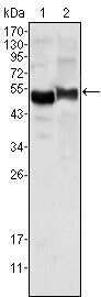
- Western blot analysis of AAT using AAT monoclonal antibody (Product # MA5-15521) in human plasma (1) and NIH/3T3 cell lysate (2).
- Submitted by
- Invitrogen Antibodies (provider)
- Main image

- Experimental details
- Western blot analysis of AAT using AAT monoclonal antibody (Product # MA5-15521) in human plasma (1) and NIH/3T3 cell lysate (2).

- Submitted by
- Invitrogen Antibodies (provider)
- Main image
- Experimental details
- Western blot analysis of AAT using AAT monoclonal antibody (Product # MA5-15521) in human plasma (1) and NIH/3T3 cell lysate (2).

- Submitted by
- Invitrogen Antibodies (provider)
- Main image

- Experimental details
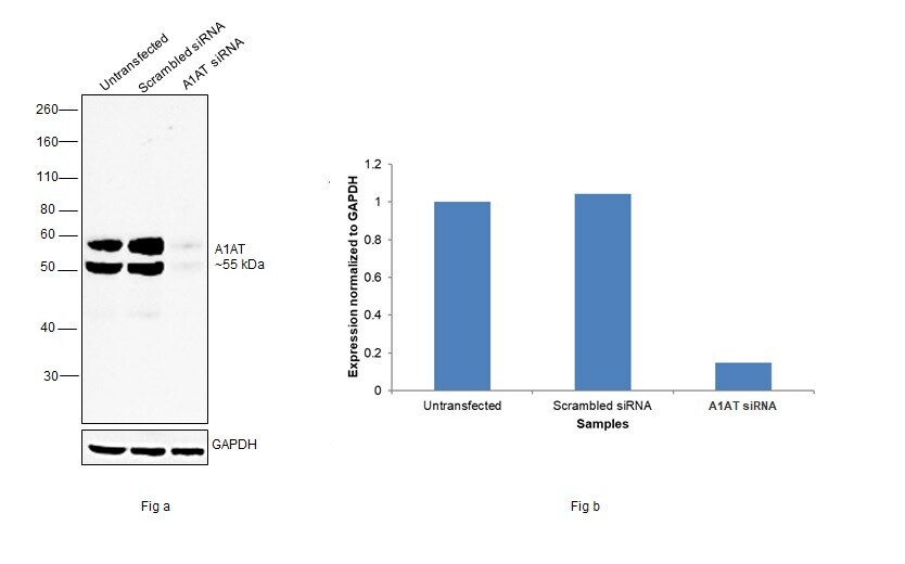
- Knockdown of alpha-1 Antitrypsin was achieved by transfecting Hep G2 with alpha-1 Antitrypsin specific siRNAs (Silencer® select Product # S10459, S10458 ). Western blot analysis (Fig. a) was performed using Whole Cell Extract-TEB from the untransfected cells (lane 1), non-targeting scrambled siRNA transfected cells (lane 2) and alpha-1 Antitrypsin knockdown cells (lane 3). The blot was probed with alpha-1 Antitrypsin Monoclonal Antibody (2B12) (Product # MA5-15521, 1:1000 dilution) and Goat anti-Mouse IgG (H+L) Superclonal™ Recombinant Secondary Antibody, HRP (Product # A28177, 1:4000 dilution). Densitometric analysis of this western blot is shown in histogram (Fig. b). Decrease in signal upon siRNA mediated knock down confirms that antibody is specific to alpha-1 Antitrypsin.
- Submitted by
- Invitrogen Antibodies (provider)
- Main image
- Experimental details
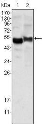
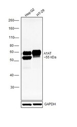
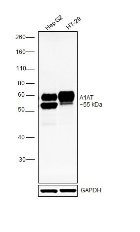
- Western blot was performed using Anti-alpha-1 Antitrypsin Monoclonal Antibody (2B12)(Product # MA5-15521) and a ~55kDa band corresponding to alpha-1 Antitrypsin was observed across cell lines. Whole Cell Extract-TEB (30 µg lysate) of Hep G2 (Lane 1) and HT-29 (Lane 2) were electrophoresed using NuPAGE™ 10% Bis-Tris Protein Gel (Product # NP0301BOX). Resolved proteins were then transferred onto a Nitrocellulose membrane (Product # IB23001) by iBlot® 2 Dry Blotting System (Product # IB21001). The blot was probed with the primary antibody (1:1000 dilution) and detected by chemiluminescence with Goat anti-Mouse IgG (H+L) Superclonal™ Recombinant Secondary Antibody, HRP (Product # A28177,1:4000 dilution) using the iBright FL 1000 (Product # A32752). Chemiluminescent detection was performed using Novex® ECL Chemiluminescent Substrate Reagent Kit (Product # WP20005).
Supportive validation
- Submitted by
- Invitrogen Antibodies (provider)
- Main image
- Experimental details
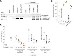
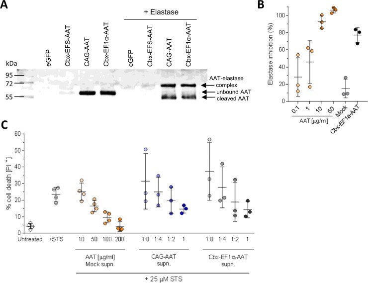
- Fig. 3 Functionality of transgenic AAT. A Formation of a complex between AAT and elastase. AAT present in the MPhi supernatant binds elastase and forms a complex represented as a band with higher molecular weight detected with anti-human AAT antibody in western blot analysis. In addition to the complex also a cleaved form of AAT of approx. 45-50 kD is visible. Same samples were used as for western blot in Fig. 1H . B Elastase inhibition by supernatant of Cbx-EF1alpha-AAT MPhi. AAT protein at different concentrations was used as a positive control. Supernatant of mock-transduced MPhi served as base for AAT protein and conditioned medium dilutions. n = 3 technical replicates. Lines represent mean +- SD. Statistical analysis was performed using one-way ANOVA with Tukey's post-hoc test. C Percentage of propidium iodide (PI) positive mAM cells after apoptosis induction with 25 uM Staurosporine (STS). AAT protein at concentrations of 10-200 ug/ml in supernatant (supn.) of mock-transduced MPhi served as positive control. Supernatants of CAG-AAT and Cbx-EF1alpha-AAT MPhi were added pure, undiluted (1) or in 1:2, 1:4, and 1:8 dilution. Untreated, +STS, AAT 10-200 mug/ml n = 4; CAG-AAT and Cbx-EF1alpha-AAT 1:8-1 n = 2 biological replicates from independent transductions and 1 technical replicate. Lines represent mean +- SD.
- Submitted by
- Invitrogen Antibodies (provider)
- Main image
- Experimental details
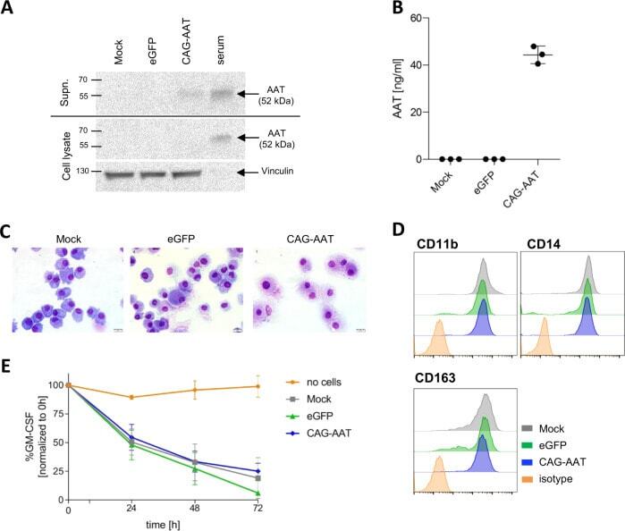
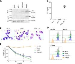
- Fig. 5 Expression of AAT in human MPhi. A Representative AAT western blot analysis of human MPhi supernatant and MPhi lysates. Human serum was used as a positive control. Vinculin band in cellular lysates served as loading control. B ELISA quantification of AAT secretion by human MPhi. n = 3. C Representative May-Grunwald-Giemsa staining of MPhi cytospins. Scale bar = 20 mum. D Representative flow cytometric analysis of myeloid and MPhi-specific surface marker expression by human MPhi. E GM-CSF uptake from cell culture medium by MPhi. A well without cells was used as a negative control. n = 3. All data represent independent cell isolations and transductions.