Explore
 Validate
 Learn
LearnAF1268
antibody from R&D Systems
Targeting: SERPINA1
A1A, A1AT, AAT, alpha-1-antitrypsin, alpha1AT, PI, PI1
 Western blot
 Immunoprecipitation
Antibody data
- Antibody Data
- Antigen structure
- References [1]
- Comments [0]
- Validations
- Western blot [4]
- Immunocytochemistry [2]
Submit
Validation data
Reference
Comment
Report error
- Product number
- AF1268 - Provider product page

- Provider
- R&D Systems
- Product name
- Human Serpin A1/alpha 1-Antitrypsin Antibody
- Antibody type
- Polyclonal
- Description
- Antigen Affinity-purified. Detects human Serpin A1/alpha 1-Antitrypsin in direct ELISAs and Western blots. In direct ELISAs, less than 5% cross-reactivity with recombinant human (rh) Serpin A3, rhSerpin A5, rhSerpin A6, rhSerpin A7, rhSerpin A11, and rhSerpin A12 is observed.
- Reactivity
- Human
- Host
- Goat
- Conjugate
- Unconjugated
- Antigen sequence
P01009- Isotype
- IgG
- Vial size
- 100 ug
- Concentration
- LYOPH
- Storage
- Use a manual defrost freezer and avoid repeated freeze-thaw cycles. 12 months from date of receipt, -20 to -70 °C as supplied. 1 month, 2 to 8 °C under sterile conditions after reconstitution. 6 months, -20 to -70 °C under sterile conditions after reconstitution.
Submitted references S-glutathionylated serine proteinase inhibitors as plasma biomarkers in assessing response to redox-modulating drugs.
Grek CL, Townsend DM, Uys JD, Manevich Y, Coker WJ 3rd, Pazoles CJ, Tew KD
Cancer research 2012 May 1;72(9):2383-93
Cancer research 2012 May 1;72(9):2383-93
No comments: Submit comment
Supportive validation
- Submitted by
- R&D Systems (provider)
- Main image

- Experimental details
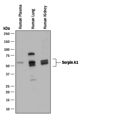
- Detection of Human Serpin A1/alpha 1-Antitrypsin by Western Blot. Western blot shows human plasma and lysates of human lung tissue and human kidney tissue. PVDF membrane was probed with 0.1 µg/mL of Goat Anti-Human Serpin A1/alpha 1-Antitrypsin Antigen Affinity-purified Polyclonal Antibody (Catalog # AF1268) followed by HRP-conjugated Anti-Goat IgG Secondary Antibody (Catalog # HAF017). Specific bands were detected for Serpin A1/alpha 1-Antitrypsin at approximately 50-60 kDa (as indicated). This experiment was conducted under reducing conditions and using Immunoblot Buffer Group 1.
- Submitted by
- R&D Systems (provider)
- Main image
- Experimental details
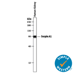
- Detection of Human Serpin A1/alpha 1-Antitrypsin by Simple WesternTM. Simple Western lane view shows lysates of human kidney tissue, loaded at 0.2 mg/mL. A specific band was detected for Serpin A1/alpha 1-Antitrypsin at approximately 65 kDa (as indicated) using 1 µg/mL of Goat Anti-Human Serpin A1/alpha 1-Antitrypsin Antigen Affinity-purified Polyclonal Antibody (Catalog # AF1268) . This experiment was conducted under reducing conditions and using the 12-230 kDa separation system.

- Submitted by
- R&D Systems (provider)
- Main image

- Experimental details
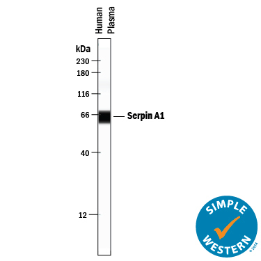
- Detection of Human Serpin A1/alpha 1-Antitrypsin by Simple WesternTM. Simple Western lane view shows human plasma, loaded at 0.2 mg/mL. A specific band was detected for Serpin A1/alpha 1-Antitrypsin at approximately 62 kDa (as indicated) using 10 µg/mL of Goat Anti-Human Serpin A1/alpha 1-Antitrypsin Antigen Affinity-purified Polyclonal Antibody (Catalog # AF1268) . This experiment was conducted under reducing conditions and using the 12-230 kDa separation system.
- Submitted by
- R&D Systems (provider)
- Main image
- Experimental details
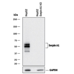
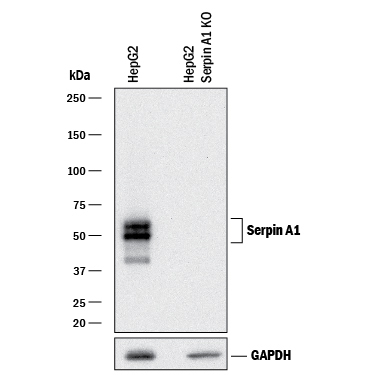
- Western Blot Shows Human Serpin A1/alpha 1-Antitrypsin Specificity by Using Knockout Cell Line. Western blot shows lysates of HepG2 human hepatocellular carcinoma parental cell line and Serpin A1/alpha 1-Antitrypsin HepG2 knockout cell line (KO). PVDF membrane was probed with 0.25 µg/mL of Goat Anti-Human Serpin A1/alpha 1-Antitrypsin Antigen Affinity-purified Polyclonal Antibody (Catalog # AF1268) followed by HRP-conjugated Anti-Goat IgG Secondary Antibody (Catalog # HAF017). Specific bands were detected for Serpin A1/alpha 1-Antitrypsin at approximately 45-55 kDa (as indicated) in the parental HepG2 cell line, but is not detectable in knockout HepG2 cell line. GAPDH (Catalog # AF5718) is shown as a loading control. This experiment was conducted under reducing conditions and using Immunoblot Buffer Group 1.
Supportive validation
- Submitted by
- R&D Systems (provider)
- Main image

- Experimental details
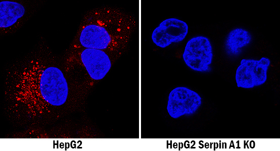
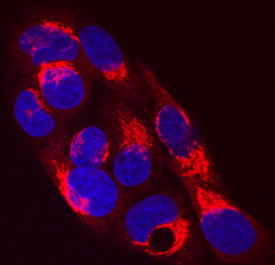
- Serpin A1/alpha 1-Antitrypsin in HepG2 Human Cell Line. Serpin A1/alpha 1-Antitrypsin was detected in immersion fixed HepG2 human hepatocellular carcinoma cell line using Goat Anti-Human Serpin A1/alpha 1-Antitrypsin Antigen Affinity-purified Polyclonal Antibody (Catalog # AF1268) at 10 µg/mL for 3 hours at room temperature. Cells were stained using the NorthernLights™ 557-conjugated Anti-Goat IgG Secondary Antibody (red; Catalog # NL001) and counterstained with DAPI (blue). Specific staining was localized to cytoplasm. View our protocol for Fluorescent ICC Staining of Cells on Coverslips.
- Submitted by
- R&D Systems (provider)
- Main image
- Experimental details
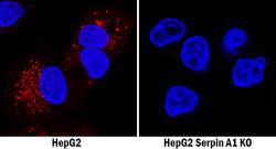
- Serpin A1/alpha 1-Antitrypsin Specificity is Shown by Immunocytochemistry in Knockout Cell Line. Serpin A1/alpha 1-Antitrypsin was detected in immersion fixed HepG2 human hepatocellular carcinoma cell line but is not detected in Serpin A1/alpha 1-Antitrypsin knockout (KO) HepG2 cell line using Goat Anti-Human Serpin A1/alpha 1-Antitrypsin Antigen Affinity-purified Polyclonal Antibody (Catalog # AF1268) at 9 µg/mL for 3 hours at room temperature. Cells were stained using the NorthernLights 557-conjugated Anti-Goat IgG Secondary Antibody (red; Catalog # NL001) and counterstained with DAPI (blue). Specific staining was localized to cytoplasm. View our protocol for Fluorescent ICC Staining of Cells on Coverslips.