 Explore
 Validate
 Learn
Learn Western blot
 ELISA
ELISAAntibody data
- Antibody Data
- Antigen structure
- References [0]
- Comments [0]
- Validations
- Western blot [2]
- Immunocytochemistry [1]
- Blocking/Neutralizing [1]
Submit
Validation data
Reference
Comment
Report error
- Product number
- MAB601R-100 - Provider product page

- Provider
- R&D Systems
- Product name
- Human IL-1 beta/IL-1F2 Antibody
- Antibody type
- Monoclonal
- Description
- Protein A or G purified from cell culture supernatant. Detects human IL-1 beta/IL-1F2 in sandwich ELISAs and Western blots. In sandwich ELISAs, less than 4% cross-reactivity with recombinant rat (rr) IL-1 beta and less than 0.1% with recombinant porcine (rp) IL-1 beta , recombinant human IL-1 alpha , rpIL-1 alpha , rrIL-1 alpha , recombinant mouse (rm) IL-1 alpha , and rmIL-1 beta is observed.
- Reactivity
- Human
- Host
- Mouse
- Conjugate
- Unconjugated
- Isotype
- IgG
- Antibody clone number
- 2805R
- Vial size
- 100 ug
- Storage
- Use a manual defrost freezer and avoid repeated freeze-thaw cycles. 12 months from date of receipt, -20 to -70 °C as supplied. 1 month, 2 to 8 °C under sterile conditions after reconstitution. 6 months, -20 to -70 °C under sterile conditions after reconstitution.
No comments: Submit comment
Supportive validation
- Submitted by
- R&D Systems (provider)
- Main image

- Experimental details
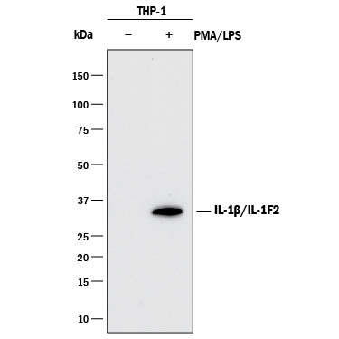
- Detection of Human IL-1 beta/IL-1F2 by Western Blot. Western blot shows lysates of THP-1 human acute monocytic leukemia cell line untreated (-) or treated (+) with 200 nM PMA for 24 hours and 10 μg/mL LPS for 3 hours. PVDF membrane was probed with 1 µg/mL of Mouse Anti-Human IL-1 beta/IL-1F2 Monoclonal Antibody (Catalog # MAB601R) followed by HRP-conjugated Anti-Mouse IgG Secondary Antibody (Catalog # HAF018). A specific band was detected for IL-1 beta/IL-1F2 at approximately 35 kDa (as indicated). This experiment was conducted under reducing conditions and using Immunoblot Buffer Group 1.
- Submitted by
- R&D Systems (provider)
- Main image
- Experimental details
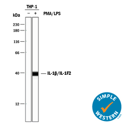
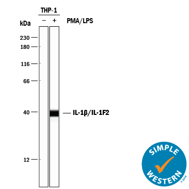
- Detection of Human IL-1 beta/IL-1F2 by Simple WesternTM. Simple Western lane view shows lysates of THP-1 human acute monocytic leukemia cell line untreated (-) or treated (+) with 200 nM PMA for 200 nM and 10 μg/mL LPS for 3 hours, loaded at 0.2 mg/mL. A specific band was detected for IL-1 beta/IL-1F2 at approximately 37 kDa (as indicated) using 10 µg/mL of Mouse Anti-Human IL-1 beta/IL-1F2 Monoclonal Antibody (Catalog # MAB601R) . This experiment was conducted under reducing conditions and using the 12-230 kDa separation system.
Supportive validation
- Submitted by
- R&D Systems (provider)
- Main image

- Experimental details
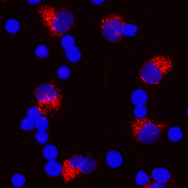
- IL-1 beta/IL-1F2 in Human PBMCs. IL-1 beta/IL-1F2 was detected in immersion fixed human peripheral blood mononuclear cells (PBMCs) treated with 1 μg/mL LPS and 3 μM monensin for 24 hours using Mouse Anti-Human IL-1 beta/IL-1F2 Monoclonal Antibody (Catalog # MAB601R) at 10 µg/mL for 3 hours at room temperature. Cells were stained using the NorthernLights™ 557-conjugated Anti-Mouse IgG Secondary Antibody (red; Catalog # NL007) and counterstained with DAPI (blue). Specific staining was localized to cytoplasm. View our protocol for Fluorescent ICC Staining of Non-adherent Cells.
Supportive validation
- Submitted by
- R&D Systems (provider)
- Main image

- Experimental details
- Cell Proliferation Induced by IL-1 beta/IL-1F2 and Neutralization by Human IL-1 beta/IL-1F2 Antibody. Recombinant HumanIL-1 beta/IL-1F2 (Catalog # 201-LB) stimulates proliferation in the the D10.G4.1 mouse helper T cell line in a dose-dependent manner (orange line) as measured by Resazurin (Catalog # AR002). Proliferation elicited by Recombinant Human IL-1 beta/IL-1F2 (50 pg/mL) is neutralized (green line) by increasing concentrations of Mouse Anti-Human IL-1 beta/IL-1F2 Monoclonal Antibody (Catalog # MAB601R). The ND50 is typically 50-200 ng/mL.
