 Explore
 Validate
 Learn
LearnR1191P
antibody from Acris Antibodies GmbH
Targeting: IL1B
IL-1B, IL1-BETA, IL1F2
 Western blot
 ELISA Immunocytochemistry Immunoprecipitation Immunohistochemistry Blocking/Neutralizing Radioimmunoassay
Antibody data
- Antibody Data
- Antigen structure
- References [0]
- Comments [0]
- Validations
- Western blot [1]
- Immunocytochemistry [1]
Submit
Validation data
Reference
Comment
Report error
- Product number
- R1191P - Provider product page

- Provider
- Acris Antibodies GmbH
- Proper citation
- Acris Antibodies GmbH Cat#R1191P, RRID:AB_10845328
- Product name
- anti Interleukin-1 beta / IL-1B
- Antibody type
- Polyclonal
- Antigen
- Recombinant Mouse IL-1 beta produced in E. coli
- Reactivity
- Mouse, Rat
- Host
- Rabbit
- Vial size
- 0.1 mg
- Concentration
- 1.0 mg/ml (UV absorbance at 280 nm)
No comments: Submit comment
Supportive validation
- Submitted by
- Acris Antibodies GmbH (provider)
- Main image

- Experimental details
- Figure 2. This antibody will recognize 10% of the non-denatured (native) precursor 31,000 MW Mouse IL-1beta containing samples but will primarily detect all of the 17,000 MW mature molecule. However, in western blot analysis, the usual procedure of heating the sample in SDS with or without reducing agents will facilitate denaturing of the 31,000 MW IL-1beta precursor molecule. Denatured IL-1beta will have a 18 kDa band.

Supportive validation
- Submitted by
- Acris Antibodies GmbH (provider)
- Main image
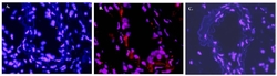
- Experimental details
- Figure 1. Immunofluorescence Microscopy after staining of Mouse carotid artery tissue with anti-Mouse IL1-beta antiserum (Less purified form of R1191P) diluted 1/50. Tissue sections were prepared after cyrofixation. Reaction occurred at RT for 60' followed by washes and reaction with Rhodamine conjugated Goat anti Rabbit IgG (R1454T). Tissue was counterstained with bis-benzimide solution at 0.5 mg/ml in PBS for 15 min at RT. Panel A) shows no antibody staining of WT uninjured mouse carotid tissue. Panel B) shows anti-IL-1beta staining of cells after surgical injury of tissue. Panel C) shows no antibody staining of injured carotid tissue from an IL-1beta KO mouse.
