Explore
 Validate
 Learn
Learn Western blot
 Immunoprecipitation
Antibody data
- Antibody Data
- Antigen structure
- References [9]
- Comments [0]
- Validations
- Western blot [2]
- Immunohistochemistry [2]
Submit
Validation data
Reference
Comment
Report error
- Product number
- AF902 - Provider product page

- Provider
- Novus Biologicals
- Product name
- Goat Polyclonal MMP-2 Antibody
- Antibody type
- Polyclonal
- Description
- Antigen Affinity-purified. Detects human MMP-2 in direct ELISAs and Western blots. In direct ELISAs, approximately 50-100% cross-reactivity with recombinant mouse MMP-2 is observed, approximately 15% cross-reactivity with recombinant human (rh) MMP-7 and less than 5% cross-reactivity with rhMMP-1, rhMMP-3, rhMMP-8, rhMMP-9, rhMMP-10, rhMMP-12, rhMMP-13, rhMMP-14, rhMMP-15, rhMMP-16, rhMMP-17, rhMMP-19, and rhMMP-24 is observed.
- Reactivity
- Human
- Host
- Goat
- Conjugate
- Unconjugated
- Isotype
- IgG
- Vial size
- 100 ug
- Concentration
- LYOPH
- Storage
- Use a manual defrost freezer and avoid repeated freeze-thaw cycles. 12 months from date of receipt, -20 to -70 degreesC as supplied. 1 month, 2 to 8 degreesC under sterile conditions after reconstitution. 6 months, -20 to -70 degreesC under sterile conditions after reconstitution.
Submitted references Glycosaminoglycans influence enzyme activity of MMP2 and MMP2/TIMP3 complex formation - Insights at cellular and molecular level.
EDTA/gelatin zymography method to identify C1s versus activated MMP-9 in plasma and immune complexes of patients with systemic lupus erythematosus.
Age-related lung tissue remodeling due to the local distribution of MMP-2, TIMP-2, TGF-β and Hsp70.
Humanin, a cytoprotective peptide, is expressed in carotid atherosclerotic [corrected] plaques in humans.
ErbB2-enhanced invasiveness of H-Ras MCF10A breast cells requires MMP-13 and uPA upregulation via p38 MAPK signaling.
Fibroblast-conditioned media promote human sarcoma cell invasion.
Development and validation of sandwich ELISA microarrays with minimal assay interference.
Claudin-1 overexpression in melanoma is regulated by PKC and contributes to melanoma cell motility.
Differential expression and activity of matrix metalloproteinases during flow-modulated vein graft remodeling.
Ruiz-Gómez G, Vogel S, Möller S, Pisabarro MT, Hempel U
Scientific reports 2019 Mar 20;9(1):4905
Scientific reports 2019 Mar 20;9(1):4905
EDTA/gelatin zymography method to identify C1s versus activated MMP-9 in plasma and immune complexes of patients with systemic lupus erythematosus.
Ugarte-Berzal E, Martens E, Boon L, Vandooren J, Blockmans D, Proost P, Opdenakker G
Journal of cellular and molecular medicine 2019 Jan;23(1):576-585
Journal of cellular and molecular medicine 2019 Jan;23(1):576-585
Age-related lung tissue remodeling due to the local distribution of MMP-2, TIMP-2, TGF-β and Hsp70.
Vitenberga Z, Pilmane M
Biotechnic & histochemistry : official publication of the Biological Stain Commission 2018;93(4):239-248
Biotechnic & histochemistry : official publication of the Biological Stain Commission 2018;93(4):239-248
Humanin, a cytoprotective peptide, is expressed in carotid atherosclerotic [corrected] plaques in humans.
Zacharias DG, Kim SG, Massat AE, Bachar AR, Oh YK, Herrmann J, Rodriguez-Porcel M, Cohen P, Lerman LO, Lerman A
PloS one 2012;7(2):e31065
PloS one 2012;7(2):e31065
ErbB2-enhanced invasiveness of H-Ras MCF10A breast cells requires MMP-13 and uPA upregulation via p38 MAPK signaling.
Yong HY, Kim IY, Kim JS, Moon A
International journal of oncology 2010 Feb;36(2):501-7
International journal of oncology 2010 Feb;36(2):501-7
Fibroblast-conditioned media promote human sarcoma cell invasion.
Bittner JG 4th, Wilson M, Shah MB, Albo D, Feig BW, Wang TN
Surgery 2009 Jan;145(1):42-7
Surgery 2009 Jan;145(1):42-7
Development and validation of sandwich ELISA microarrays with minimal assay interference.
Gonzalez RM, Seurynck-Servoss SL, Crowley SA, Brown M, Omenn GS, Hayes DF, Zangar RC
Journal of proteome research 2008 Jun;7(6):2406-14
Journal of proteome research 2008 Jun;7(6):2406-14
Claudin-1 overexpression in melanoma is regulated by PKC and contributes to melanoma cell motility.
Leotlela PD, Wade MS, Duray PH, Rhode MJ, Brown HF, Rosenthal DT, Dissanayake SK, Earley R, Indig FE, Nickoloff BJ, Taub DD, Kallioniemi OP, Meltzer P, Morin PJ, Weeraratna AT
Oncogene 2007 May 31;26(26):3846-56
Oncogene 2007 May 31;26(26):3846-56
Differential expression and activity of matrix metalloproteinases during flow-modulated vein graft remodeling.
Berceli SA, Jiang Z, Klingman NV, Pfahnl CL, Abouhamze ZS, Frase CD, Schultz GS, Ozaki CK
Journal of vascular surgery 2004 May;39(5):1084-90
Journal of vascular surgery 2004 May;39(5):1084-90
No comments: Submit comment
Supportive validation
- Submitted by
- Novus Biologicals (provider)
- Main image
- Experimental details
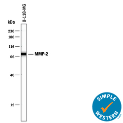
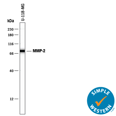
- Detection of Human MMP-2 by Simple WesternTM. Simple Western lane view shows lysates of U-118-MG human glioblastoma/astrocytoma cell line, loaded at 0.2 mg/mL. A specific band was detected for MMP-2 at approximately 78 kDa (as indicated) using 10 µg/mL of Goat Anti-Human MMP-2 Antigen Affinity-purified Polyclonal Antibody (Catalog # AF902) followed by 1:50 dilution of HRP-conjugated Anti-Goat IgG Secondary Antibody (Catalog # HAF109). This experiment was conducted under reducing conditions and using the 12-230 kDa separation system.
- Submitted by
- Novus Biologicals (provider)
- Main image

- Experimental details
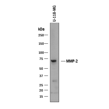
- Detection of Human MMP-2 by Western Blot. Western blot shows lysate of U-118-MG human glioblastoma/astrocytoma cell line. PVDF membrane was probed with 1 µg/mL of Goat Anti-Human MMP-2 Antigen Affinity-purified Polyclonal Antibody (Catalog # AF902) followed by HRP-conjugated Anti-Goat IgG Secondary Antibody (Catalog # HAF017). A specific band was detected for MMP-2 at approximately 72 kDa (as indicated). This experiment was conducted under reducing conditions and using Immunoblot Buffer Group 1.
Supportive validation
- Submitted by
- Novus Biologicals (provider)
- Main image
- Experimental details
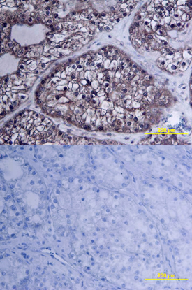
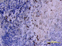
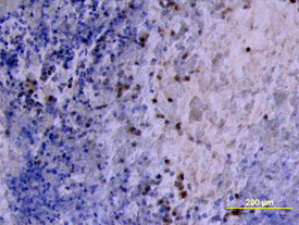
- MMP-2 in Human Ovarian Cancer Tissue. MMP-2 was detected in immersion fixed paraffin-embedded sections of human ovarian cancer tissue using Goat Anti-Human MMP-2 Antigen Affinity-purified Poly-clonal Antibody (Catalog # AF902) at 10 µg/mL overnight at 4 °C. Tissue was stained using the Anti-Goat HRP-DAB Cell & Tissue Staining Kit (brown; Catalog # CTS008) and counter-stained with hematoxylin (blue). View our protocol for Chromogenic IHC Staining of Paraffin-embedded Tissue Sections.
- Submitted by
- Novus Biologicals (provider)
- Main image
- Experimental details
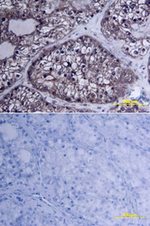
- MMP-2 in Human Ovary. MMP-2 was detected in immersion fixed paraffin-embedded sections of human ovarian array using Goat Anti-Human MMP-2 Antigen Affinity-purified Polyclonal Antibody (Catalog # AF902) at 10 µg/mL overnight at 4 °C. Tissue was stained using the Anti-Goat HRP-DAB Cell & Tissue Staining Kit (brown; Catalog # CTS008) and counterstained with hematoxylin (blue). Lower panel shows a lack of labeling if primary antibodies are omitted and tissue is stained only with secondary antibody followed by incubation with detection reagents. View our protocol for Chromogenic IHC Staining of Paraffin-embedded Tissue Sections.