Explore
 Validate
 Learn
Learn Western blot
 ELISA
ELISAAntibody data
- Antibody Data
- Antigen structure
- References [4]
- Comments [0]
- Validations
- Western blot [2]
- Immunohistochemistry [3]
- Flow cytometry [1]
Submit
Validation data
Reference
Comment
Report error
- Product number
- NB200-113 - Provider product page

- Provider
- Novus Biologicals
- Proper citation
- Novus Cat#NB200-113, RRID:AB_10000526
- Product name
- Mouse Monoclonal MMP-2 Antibody
- Antibody type
- Monoclonal
- Description
- Protein G purified. This is specific for pro and active MMP-2.
- Reactivity
- Human, Mouse, Rat, Guinea Pig
- Host
- Mouse
- Isotype
- IgG
- Vial size
- 0.1 ml
- Concentration
- 1.0 mg/ml
- Storage
- Aliquot and store at -20C or -80C. Avoid freeze-thaw cycles.
Submitted references Adipose-derived stem cells delay muscle atrophy after peripheral nerve injury in the rodent model.
Effects of acupuncture at GV20 and ST36 on the expression of matrix metalloproteinase 2, aquaporin 4, and aquaporin 9 in rats subjected to cerebral ischemia/reperfusion injury.
Exercise-induced regulation of matrix metalloproteinases in the skeletal muscle of subjects with type 2 diabetes.
Acetaminophen attenuates peroxynitrite-activated matrix metalloproteinase-2-mediated troponin I cleavage in the isolated guinea pig myocardium.
Schilling BK, Schusterman MA 2nd, Kim DY, Repko AJ, Klett KC, Christ GJ, Marra KG
Muscle & nerve 2019 May;59(5):603-610
Muscle & nerve 2019 May;59(5):603-610
Effects of acupuncture at GV20 and ST36 on the expression of matrix metalloproteinase 2, aquaporin 4, and aquaporin 9 in rats subjected to cerebral ischemia/reperfusion injury.
Xu H, Zhang Y, Sun H, Chen S, Wang F
PloS one 2014;9(5):e97488
PloS one 2014;9(5):e97488
Exercise-induced regulation of matrix metalloproteinases in the skeletal muscle of subjects with type 2 diabetes.
Scheede-Bergdahl C, Bergdahl A, Schjerling P, Qvortrup K, Koskinen SO, Dela F
Diabetes & vascular disease research 2014 Sep;11(5):324-34
Diabetes & vascular disease research 2014 Sep;11(5):324-34
Acetaminophen attenuates peroxynitrite-activated matrix metalloproteinase-2-mediated troponin I cleavage in the isolated guinea pig myocardium.
Rork TH, Hadzimichalis NM, Kappil MA, Merrill GF
Journal of molecular and cellular cardiology 2006 Apr;40(4):553-61
Journal of molecular and cellular cardiology 2006 Apr;40(4):553-61
No comments: Submit comment
Supportive validation
- Submitted by
- Novus Biologicals (provider)
- Main image
- Experimental details
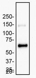
- Western Blot: MMP-2 Antibody (2C1) - (Pro and Active) [NB200-113] - Recombinant human MMP-2 expressed from CHO cells was was separated on a 7.5 % gel by SDS-PAGE, transferred to PVDF membrane and blocked in 5% non-fat milk in TBST. The membrane was probed with 2.0 ug/ml anti-MMP-2 in blocking buffer and detected with an anti-mouse HRP secondary antibody using chemiluminescence.

- Submitted by
- Novus Biologicals (provider)
- Main image
- Experimental details
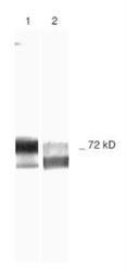
- Western Blot: MMP-2 Antibody (2C1) - (Pro and Active) [NB200-113] - Samples: 1) Human Recombinant MMP-2, and 2)Schwann Cell Culture Medium Run on 10% SDS-PAGE, followed by electroblot to nitrocellulose and slot blot immunolabeling. Antibodies: primary - NB200-113, secondary - anti-mouse-biotin. Detection: avidin-HRP. Chromagen: DAB/H202.

Supportive validation
- Submitted by
- Novus Biologicals (provider)
- Main image

- Experimental details
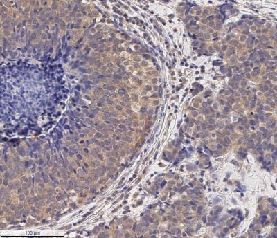
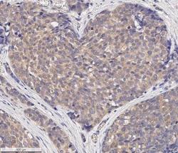
- Immunohistochemistry-Paraffin: MMP-2 Antibody (2C1) - (Pro and Active) [NB200-113] - Analysis of a FFPE human breast cancer section using 5 ug/ml conc. of MMP2 antibody (clone 2C1) on a Bond Rx autostainer (Leica Biosystems). The assay involved 20 minutes of heat induced antigen retrieval (HIER) with 10mM sodium citrate buffer (pH 6.0) and endogenous peroxidase quenching using peroxide block. The sections were incubated with primary antibody for 30 minutes. Bond Polymer Refine Detection (Leica Biosystems) and DAB were used for signal detection which followed counterstaining with hematoxylin. Whole slide scanning and capturing of representative images (20X) were performed using Aperio AT2 (Leica Biosystems). MMP2 immunoreactivity was observed in the cytoplasm of the cancer cells and the stromal cells, and in the inter-cellular spaces (secreted MMP2). The cancer cells present in the core of tumor areas were found to be negative for MMP2 staining.
- Submitted by
- Novus Biologicals (provider)
- Main image
- Experimental details
- Immunohistochemistry-Paraffin: MMP-2 Antibody (2C1) - (Pro and Active) [NB200-113] - Analysis of a FFPE human breast cancer section using 5 ug/ml conc. of MMP2 antibody (clone 2C1) on a Bond Rx autostainer (Leica Biosystems). The assay involved 20 minutes of heat induced antigen retrieval (HIER) with 10mM sodium citrate buffer (pH 6.0) and endogenous peroxidase quenching using peroxide block. The sections were incubated with primary antibody for 30 minutes. Bond Polymer Refine Detection (Leica Biosystems) and DAB were used for signal detection which followed counterstaining with hematoxylin. Whole slide scanning and capturing of representative images (20X) were performed using Aperio AT2 (Leica Biosystems). This MMP2 antibody generated an expected immunoreactivity of MMP2 protein in the cytoplasm of most of the cancer cells and the stromal cells, and in the inter-cellular spaces (secreted pool of MMP2). Staining was performed by Histowiz.

- Submitted by
- Novus Biologicals (provider)
- Main image
- Experimental details
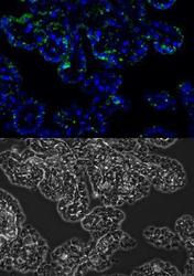
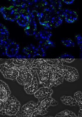
- Immunohistochemistry-Paraffin: MMP-2 Antibody (2C1) - (Pro and Active) [NB200-113] - Upper image: MMP-2 FITC (green), DAPI (blue) Lower image: phase contrast of human choroid plexus (Alzheimer's disease). Image from verified customer review.
Supportive validation
- Submitted by
- Novus Biologicals (provider)
- Main image

- Experimental details
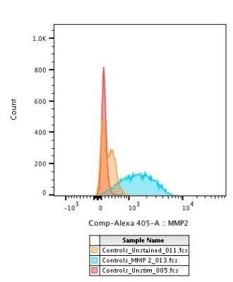
- Flow Cytometry: MMP-2 Antibody (2C1) - (Pro and Active) [NB200-113] - Flow Cytometry: MMP-2 Antibody (2C1) [Alexa Fluor® 405] [NB200-113AF405] - CD4+ T cells were activated for 24h before being fixed and permeabilized to detect MMP-2. Image using the Alexa Fluor 405 form of this antibody. Image from verified customer review.