Explore
 Validate
 Learn
LearnMA5-60184
antibody from Invitrogen Antibodies
Targeting: BRCA2
BRCC2, FACD, FAD, FAD1, FANCD, FANCD1, XRCC11
 Western blot
Antibody data
- Antibody Data
- Antigen structure
- References [0]
- Comments [0]
- Validations
- Western blot [4]
- Immunocytochemistry [1]
- Immunohistochemistry [2]
- Flow cytometry [1]
Submit
Validation data
Reference
Comment
Report error
- Product number
- MA5-60184 - Provider product page

- Provider
- Invitrogen Antibodies
- Product name
- BRCA2 Recombinant Rabbit Monoclonal Antibody (PSH07-55)
- Antibody type
- Monoclonal
- Antigen
- Recombinant full-length protein
- Description
- Positive Control: HeLa cell lysate, 293T cell lysate, Jurkat cell lysate, MCF7. Tissue Specificity: Tissue enhanced (bone marrow, lymphoid tissue, testis). Subcellular Location: Nucleus, Cytoplasm, cytoskeleton, microtubule organizing center, centrosome. Sequence Similarities: 60% Mouse/Rat. Predicted band size: 384 kDa.
- Reactivity
- Human
- Host
- Rabbit
- Isotype
- IgG
- Antibody clone number
- PSH07-55
- Vial size
- 100 μL
- Concentration
- 1 mg/mL
- Storage
- Store at 4°C short term. For long term storage, store at -20°C, avoiding freeze/thaw cycles.
No comments: Submit comment
Supportive validation
- Submitted by
- Invitrogen Antibodies (provider)
- Main image
- Experimental details
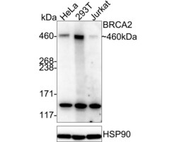
- Western blot was performed using BRCA2 Recombinant Rabbit Monoclonal Antibody (PSH07-55) (Product # MA5-60184) and 460 kDa band corresponding to BRCA2 was observed across cell lines tested. Whole cell extracts (20 µg lysate) of HeLa (Lane 1), 293T (Lane 2) and Jurkat (Lane 3) were electrophoresed using NuPAGE™ Tris-Acetate Mini Protein Gels, 3 to 8%, 1.0–1.5 mm (Proudct # EA03785BOX). Resolved proteins were transferred onto a PVDF membrane. The blot was blocked with 5% NFDM/TBST for 1 hour at room temperature, then probed with the primary antibody at 1:1,000 dilution at 4 degrees Celsius overnight and detected by chemiluminescence with HRP labeled Goat anti-Rabbit IgG secondary antibody (HA1001).

- Submitted by
- Invitrogen Antibodies (provider)
- Main image
- Experimental details
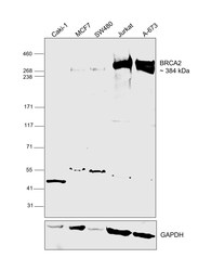
- Western blot was performed using BRCA2 Recombinant Rabbit Monoclonal Antibody (PSH07-55) (Product # MA5-60184) and a ~384 kDa band corresponding to BRCA2 was observed in all the cell lines except for Caki-1, which is a BRCA2 low expressing cell line. Whole cell extracts (60 µg) of Caki-1 (Lane 1), MCF7 (Lane 2), SW480 (Lane 3), Jurkat (Lane 4) and A-673 (Lane 5) were electrophoresed using NuPAGE™ 3-8% Tris-Acetate Protein Gel (Product # EA0378BOX). Resolved proteins were transferred onto a nitrocellulose membrane (Product # IB33001) by iBlot™ 3 Western Blot Transfer Device (Product # IB31001). The blot was probed with primary antibody (1:1,000 dilution) and detected by chemiluminescence with Goat anti-Rabbit IgG (H+L) Superclonal™ Recombinant Secondary Antibody, HRP (Product # A27036,1:20,000 dilution) using the iBright™ FL1500 Imaging System (Product # A44115). Chemiluminescent detection was performed using SuperSignal™ West Atto Ultimate Sensitivity Substrate (Product # A38555). An uncharacterized band was observed at ~120 kDa.

- Submitted by
- Invitrogen Antibodies (provider)
- Main image
- Experimental details
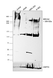
- Western blot was performed using BRCA2 Recombinant Rabbit Monoclonal Antibody (PSH07-55) (Product # MA5-60184) and a ~384 kDa band corresponding to BRCA2 was observed in all the cell lines. Whole cell extracts (60 µg) of Jurkat (Lane 1), A549 (Lane 2), HEK-293 (Lane 3) and K-562 (Lane 4) were electrophoresed using NuPAGE™ 3-8% Tris-Acetate Protein Gel (Product # EA0378BOX). Resolved proteins were transferred onto a nitrocellulose membrane (Product # IB33001) by iBlot™ 3 Western Blot Transfer Device (Product # IB31001). The blot was probed with primary antibody (1:1,000 dilution) and detected by chemiluminescence with Goat anti-Rabbit IgG (H+L) Superclonal™ Recombinant Secondary Antibody, HRP (Product # A27036,1:20,000 dilution) using the iBright™ FL1500 Imaging System (Product # A44115). Chemiluminescent detection was performed using SuperSignal™ West Atto Ultimate Sensitivity Substrate (Product # A38555). An uncharacterized band was observed at ~120 kDa.

- Submitted by
- Invitrogen Antibodies (provider)
- Main image
- Experimental details
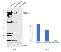
- Knockdown of BRCA2 was achieved by transfecting A-673 with BRCA2 specific siRNAs (Silencer® select Product #s2083, s2085). Western blot analysis (Fig. a) was performed using whole cell extracts from the BRCA2 knockdown cells (lane 3), non-targeting scrambled siRNA transfected cells (lane 2) and untransfected cells (lane 1). The blot was probed with BRCA2 Recombinant Rabbit Monoclonal Antibody (PSH07-55) (Product # MA5-60184), 1:1,000 dilution) and Goat anti-Rabbit IgG (H+L) Superclonal™ Recombinant Secondary Antibody, HRP (Product # A27036, 1:20,000 dilution) and detected using the iBright™ FL1500 Imaging System (Product # A44115). Densitometric analysis of this western blot is shown in histogram (Fig. b). Decrease in signal upon siRNA mediated knock down confirms that antibody is specific to BRCA2. An uncharacterized band was observed at ~120 kDa.

Supportive validation
- Submitted by
- Invitrogen Antibodies (provider)
- Main image
- Experimental details
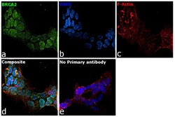
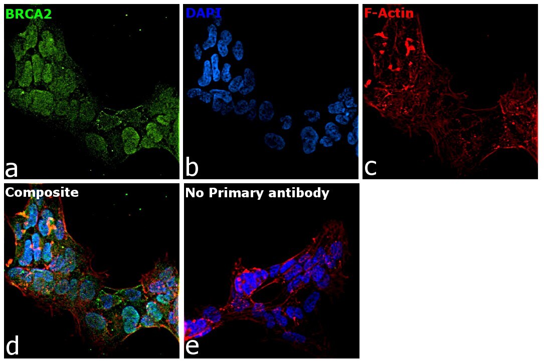
- Immunofluorescence analysis of BRCA2 was performed using 70% confluent log phase A-673 cells. The cells were fixed with 4% paraformaldehyde for 10 minutes and blocked with 2% BSA for 1 hour at room temperature. The cells were labeled with BRCA2 Recombinant Rabbit Monoclonal Antibody (PSH07-55) (Product # MA5-60184) at 1:100 dilution in 0.1% BSA, incubated at 4 degree Celsius overnight and then labeled with Donkey anti-Rabbit IgG (H+L) Highly Cross-Adsorbed Secondary Antibody, Alexa Fluor Plus 488 (Product # A32790), (1:2,000 dilution), for 45 minutes at room temperature (Panel a: Green). Nuclei (Panel b:Blue) were stained with ProLong™ Diamond Antifade Mountant with DAPI (Product # P36962). F-actin (Panel c: Red) was stained with Rhodamine Phalloidin (Product # R415, 1:300 dilution). Panel d represents the merged image showing nuclear and cytoplasmic localization. Panel e represents control cells with no primary antibody to assess background. The images were captured at 40X magnification.
Supportive validation
- Submitted by
- Invitrogen Antibodies (provider)
- Main image
- Experimental details
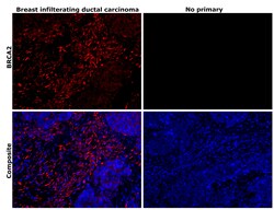
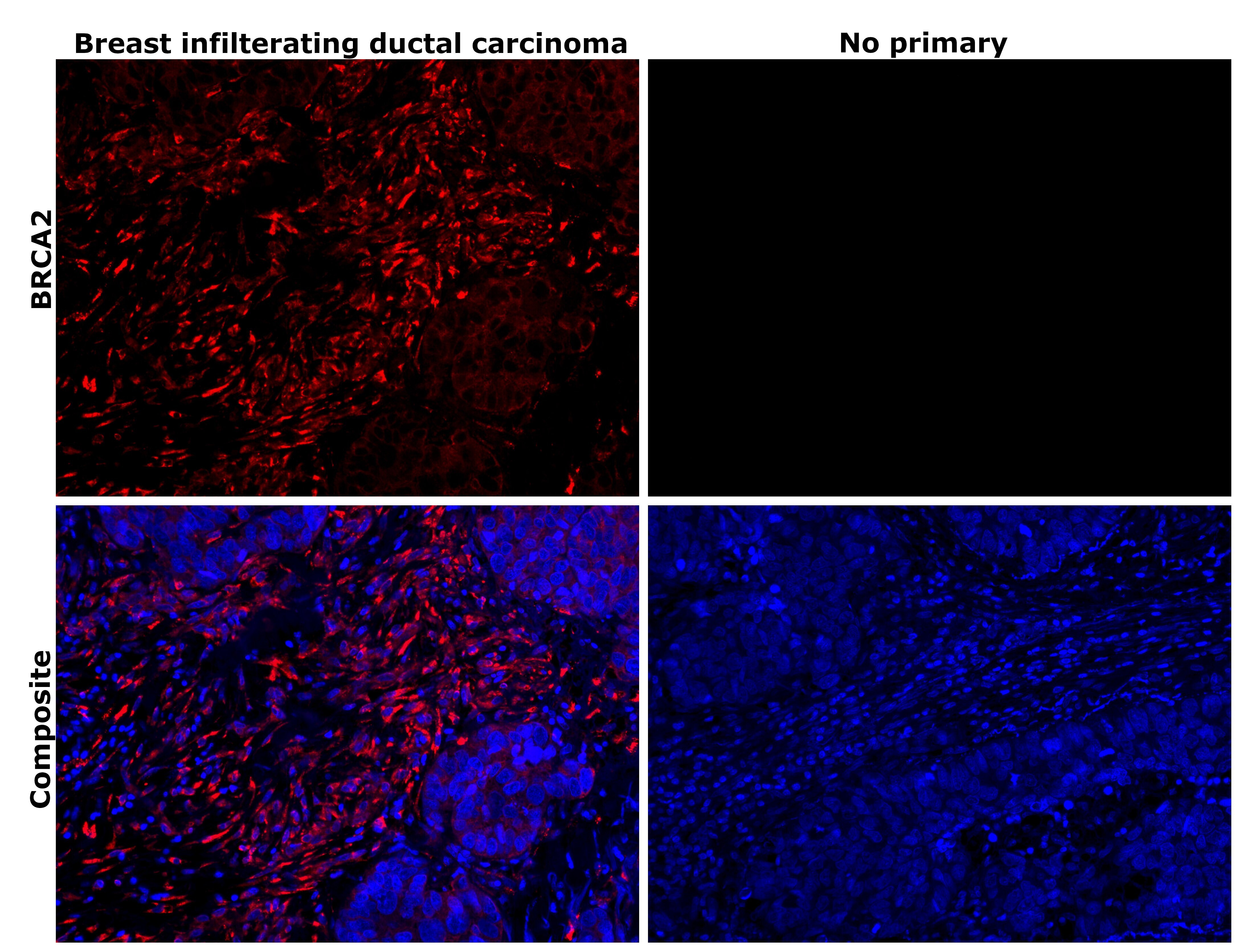
- Immunohistochemical analysis of BRCA2 was performed using formalin-fixed paraffin-embedded human breast infilterating ductal carcinoma tissue sections. To expose the target protein, heat-induced epitope retrieval was performed on de-paraffinized sections using eBioscience™ IHC Antigen Retrieval Solution - High pH (10X) (Product # 00-4956-58) diluted to 1X solution in water in a decloaking chamber at 110 degree Celsius for 15 minutes. Following antigen retrieval, the sections were blocked with 3% H2O2 for 1 hour at room temperature followed by 2% normal goat serum in 1X PBS for 45 minutes at room temperature. The sections were then probed with BRCA2 Recombinant Rabbit Monoclonal Antibody (PSH07-55) (Product # MA5-60184) at 1:500 dilution in 0.1% normal goat serum overnight at 4 degree Celsius in a humidified chamber. Detection was performed using Alexa Fluor™ 594 Tyramide SuperBoost™ Kit, goat anti-rabbit IgG (Product # B40925). Nuclei were stained with DAPI (Product # D1306) and the sections were mounted using ProLong™ Glass Antifade Mountant (Product # P36984). The images were captured on EVOS™ M7000 Imaging System (Product # AMF7000) at 20X magnification. Cytoplasmic staining pattern was observed in the cells of the tissue section.
- Submitted by
- Invitrogen Antibodies (provider)
- Main image
- Experimental details
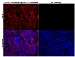
- Immunohistochemical analysis of BRCA2 was performed using formalin-fixed paraffin-embedded human breast infilterating ductal carcinoma tissue sections. To expose the target protein, heat-induced epitope retrieval was performed on de-paraffinized sections using eBioscience™ IHC Antigen Retrieval Solution - High pH (10X) (Product # 00-4956-58) diluted to 1X solution in water in a decloaking chamber at 110 degree Celsius for 15 minutes. Following antigen retrieval, the sections were blocked with 3% H2O2 for 1 hour at room temperature followed by 2% normal goat serum in 1X PBS for 45 minutes at room temperature. The sections were then probed with BRCA2 Recombinant Rabbit Monoclonal Antibody (PSH07-55) (Product # MA5-60184) at 1:500 dilution in 0.1% normal goat serum overnight at 4 degree Celsius in a humidified chamber. Detection was performed using Alexa Fluor™ 594 Tyramide SuperBoost™ Kit, goat anti-rabbit IgG (Product # B40925). Nuclei were stained with DAPI (Product # D1306) and the sections were mounted using ProLong™ Glass Antifade Mountant (Product # P36984). The images were captured on EVOS™ M7000 Imaging System (Product # AMF7000) at 20X magnification. Cytoplasmic staining pattern was observed in the cells of the tissue section.

Supportive validation
- Submitted by
- Invitrogen Antibodies (provider)
- Main image
- Experimental details
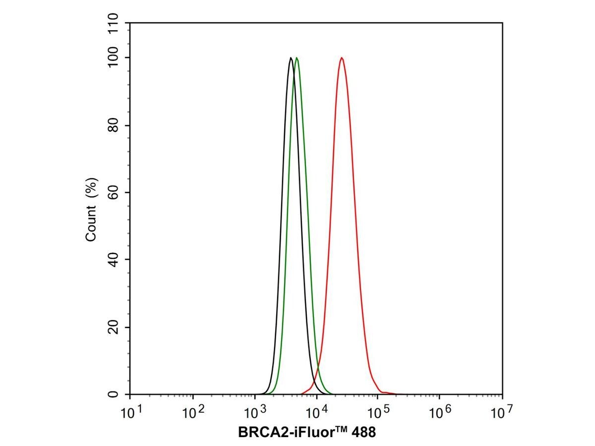
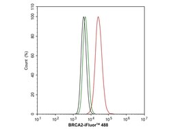
- MCF7 cells were fixed and permeabilized, and then stained with BRCA2 Recombinant Rabbit Monoclonal Antibody (PSH07-55) (Product # MA5-60184) at 1:1,000 dilution (red) or with Rabbit IgG Isotype Control (green). After incubation of the primary antibody at 4 degrees Celsius for an hour, the cells were stained with a iFluor™ 488 conjugate-Goat anti-Rabbit IgG Secondary antibody at 1:1,000 dilution for 30 minutes at 4 degrees Celsius. Unlabelled sample was used as a control (cells without incubation with primary antibody; black).