Explore
 Validate
 Learn
Learn Western blot
 Immunocytochemistry
Antibody data
- Antibody Data
- Antigen structure
- References [0]
- Comments [0]
- Validations
- Western blot [3]
- Immunocytochemistry [1]
Submit
Validation data
Reference
Comment
Report error
- Product number
- GTX108627 - Provider product page

- Provider
- GeneTex
- Proper citation
- GeneTex Cat#GTX108627, RRID:AB_10731582
- Product name
- ZO-1 antibody [N2C1], Internal
- Antibody type
- Polyclonal
- Reactivity
- Human
- Host
- Rabbit
No comments: Submit comment
Enhanced validation
Supportive validation
- Submitted by
- GeneTex (provider)
- Enhanced method
- Genetic validation
- Main image

- Experimental details
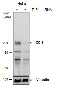
- Non-transfected (¡V) and transfected (+) HeLa whole cell extracts (30 ?g) were separated by 5% SDS-PAGE, and the membrane was blotted with ZO-1 antibody [N2C1], Internal (GTX108627) diluted at 1:500. The HRP-conjugated anti-rabbit IgG antibody (GTX213110-01) was used to detect the primary antibody.
Supportive validation
- Submitted by
- GeneTex (provider)
- Main image
- Experimental details
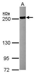
- Sample (30 ?g of whole cell lysate) A: HCT116 5% SDS PAGE GTX108627 diluted at 1:5000 The HRP-conjugated anti-rabbit IgG antibody (GTX213110-01) was used to detect the primary antibody.

- Submitted by
- GeneTex (provider)
- Main image

- Experimental details
- Non-transfected (¡V) and transfected (+) HeLa whole cell extracts (30 ?g) were separated by 5% SDS-PAGE, and the membrane was blotted with ZO-1 antibody [N2C1], Internal (GTX108627) diluted at 1:500. The HRP-conjugated anti-rabbit IgG antibody (GTX213110-01) was used to detect the primary antibody.

Supportive validation
- Submitted by
- GeneTex (provider)
- Main image
- Experimental details
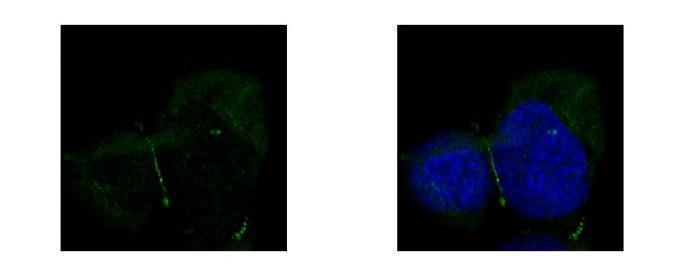
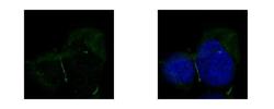
- ZO-1 antibody [N2C1], Internal detects TJP1 protein at junction by confocal immunofluorescent analysis. Sample: A431 cells were fixed in ice-cold MeOH for 5 min.Green: TJP1 protein stained by ZO-1 antibody [N2C1], Internal (GTX108627) diluted at 1:500.Blue: Hoechst 33342 staining.[Images captured by Olympus FV10i Confocal Laser Scanning Microscope]