Explore
 Validate
 Learn
Learn Western blot
 Immunocytochemistry
Antibody data
- Antibody Data
- Antigen structure
- References [35]
- Comments [0]
- Validations
- Immunocytochemistry [1]
- Other assay [31]
Submit
Validation data
Reference
Comment
Report error
- Product number
- 33-9111 - Provider product page

- Provider
- Invitrogen Antibodies
- Product name
- ZO-1 Monoclonal Antibody (ZO1-1A12), FITC
- Antibody type
- Monoclonal
- Antigen
- Recombinant full-length protein
- Description
- Species reactivity includes human (Caco-2 Cell line) and dog (MDCK cell line). Based on sequence homology, reactivity with other species is likely but has not been confirmed. This monoclonal antibody detects ZO-1alpha+ and ZO-1alpha- isoforms. The antibody is specific for ZO-1 proteins; cross-reactivity with the related ZO-2 protein has not been observed. For this FITC conjugate the excitation (EX)/emission (EM) are 494/520 (nm).
- Reactivity
- Human, Canine
- Host
- Mouse
- Conjugate
- Green dye
- Isotype
- IgG
- Antibody clone number
- ZO1-1A12
- Vial size
- 100 µg
- Concentration
- 0.5 mg/mL
- Storage
- 4° C, store in dark
Submitted references An oxygen-adaptive interaction between SNHG12 and occludin maintains blood-brain barrier integrity.
Choroidal congestion mouse model: Could it serve as a pachychoroid model?
Higher Prevalence of Bacteroides fragilis in Crohn's Disease Exacerbations and Strain-Dependent Increase of Epithelial Resistance.
Myeloid-derived suppressor cells improve corneal graft survival through suppressing angiogenesis and lymphangiogenesis.
Asymmetric Stratification-Induced Polarity Loss and Coordinated Individual Cell Movements Drive Directional Migration of Vertebrate Epithelium.
Multiorgan microfluidic platform with breathable lung chamber for inhalation or intravenous drug screening and development.
Cdh2 coordinates Myosin-II dependent internalisation of the zebrafish neural plate.
Endothelial cell rearrangements during vascular patterning require PI3-kinase-mediated inhibition of actomyosin contractility.
Design and demonstration of a pumpless 14 compartment microphysiological system.
Alix-mediated assembly of the actomyosin-tight junction polarity complex preserves epithelial polarity and epithelial barrier.
The retinal phenotype of Grk1-/- is compromised by a Crb1 rd8 mutation.
EB1 regulates tubulin and actin cytoskeletal networks at the sertoli cell blood-testis barrier in male rats: an in vitro study.
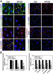
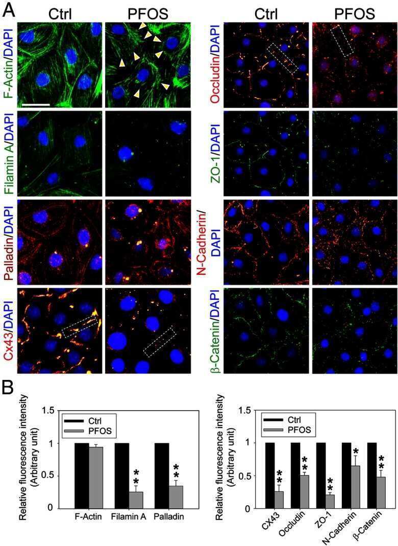
Perfluorooctanesulfonate (PFOS) perturbs male rat Sertoli cell blood-testis barrier function by affecting F-actin organization via p-FAK-Tyr(407): an in vitro study.
A multiplex high-throughput gene expression assay to simultaneously detect disease and functional markers in induced pluripotent stem cell-derived retinal pigment epithelium.
Mesoderm is required for coordinated cell movements within zebrafish neural plate in vivo.
Rai14 (retinoic acid induced protein 14) is involved in regulating f-actin dynamics at the ectoplasmic specialization in the rat testis*.
A novel human endogenous retroviral protein inhibits cell-cell fusion.
Palladin is a regulator of actin filament bundles at the ectoplasmic specialization in adult rat testes.
Tumor necrosis factor α-mediated restructuring of the Sertoli cell barrier in vitro involves matrix metalloprotease 9 (MMP9), membrane-bound intercellular adhesion molecule-1 (ICAM-1) and the actin cytoskeleton.
A mouse model for interstitial cystitis/painful bladder syndrome based on APF inhibition of bladder epithelial repair: a pilot study.
Hailey-Hailey disease and tight junctions: Claudins 1 and 4 are regulated by ATP2C1 gene encoding Ca(2+) /Mn(2+) ATPase SPCA1 in cultured keratinocytes.
Midbody accumulation through evasion of autophagy contributes to cellular reprogramming and tumorigenicity.
Protective effects of nonionic triblock copolymers on bile acid-mediated epithelial barrier disruption.
Vascular endothelial cells cultured from patients with cerebral or uncomplicated malaria exhibit differential reactivity to TNF.
Interleukin-1alpha is a regulator of the blood-testis barrier.
Adjudin-mediated Sertoli-germ cell junction disassembly affects Sertoli cell barrier function in vitro and in vivo.
Multipotent mesenchymal stem cells from human placenta: critical parameters for isolation and maintenance of stemness after isolation.
Effects of di(2-ethylhexyl) phthalate on gap and tight junction protein expression in the testis of prepubertal rats.
Interleukin 1 alpha (IL1A) is a novel regulator of the blood-testis barrier in the rat.
Localization of semaphorin 3A in the rat cornea.
Effect of low fluence diode laser irradiation on the hydraulic conductivity of perfused trabecular meshwork endothelial cell monolayers.
Dynamin II interacts with the cadherin- and occludin-based protein complexes at the blood-testis barrier in adult rat testes.
Disruption of Sertoli-germ cell adhesion function in the seminiferous epithelium of the rat testis can be limited to adherens junctions without affecting the blood-testis barrier integrity: an in vivo study using an androgen suppression model.
Apical localization of a functional TRPC3/TRPC6-Ca2+-signaling complex in polarized epithelial cells. Role in apical Ca2+ influx.
Adhering junction dynamics in the testis are regulated by an interplay of beta 1-integrin and focal adhesion complex-associated proteins.
Li Y, Wei JY, Liu H, Wang KJ, Jin SN, Su ZK, Wang HJ, Shi JX, Li B, Shang DS, Fang WG, Qin XX, Zhao WD, Chen YH
Cell reports 2022 Apr 12;39(2):110656
Cell reports 2022 Apr 12;39(2):110656
Choroidal congestion mouse model: Could it serve as a pachychoroid model?
Matsumoto H, Mukai R, Hoshino J, Oda M, Matsuzaki T, Ishizaki Y, Shibasaki K, Akiyama H
PloS one 2021;16(1):e0246115
PloS one 2021;16(1):e0246115
Higher Prevalence of Bacteroides fragilis in Crohn's Disease Exacerbations and Strain-Dependent Increase of Epithelial Resistance.
Becker HEF, Jamin C, Bervoets L, Boleij A, Xu P, Pierik MJ, Stassen FRM, Savelkoul PHM, Penders J, Jonkers DMAE
Frontiers in microbiology 2021;12:598232
Frontiers in microbiology 2021;12:598232
Myeloid-derived suppressor cells improve corneal graft survival through suppressing angiogenesis and lymphangiogenesis.
Ren Y, Dong X, Zhao H, Feng J, Chen B, Zhou Y, Peng Y, Zhang L, Zhou Q, Li Y, Wu M, He Y
American journal of transplantation : official journal of the American Society of Transplantation and the American Society of Transplant Surgeons 2021 Feb;21(2):552-566
American journal of transplantation : official journal of the American Society of Transplantation and the American Society of Transplant Surgeons 2021 Feb;21(2):552-566
Asymmetric Stratification-Induced Polarity Loss and Coordinated Individual Cell Movements Drive Directional Migration of Vertebrate Epithelium.
Lu Y, Deng R, You H, Xu Y, Antos C, Sun J, Klein OD, Lu P
Cell reports 2020 Oct 13;33(2):108246
Cell reports 2020 Oct 13;33(2):108246
Multiorgan microfluidic platform with breathable lung chamber for inhalation or intravenous drug screening and development.
Miller PG, Chen CY, Wang YI, Gao E, Shuler ML
Biotechnology and bioengineering 2020 Feb;117(2):486-497
Biotechnology and bioengineering 2020 Feb;117(2):486-497
Cdh2 coordinates Myosin-II dependent internalisation of the zebrafish neural plate.
Araya C, Häkkinen HM, Carcamo L, Cerda M, Savy T, Rookyard C, Peyriéras N, Clarke JDW
Scientific reports 2019 Feb 12;9(1):1835
Scientific reports 2019 Feb 12;9(1):1835
Endothelial cell rearrangements during vascular patterning require PI3-kinase-mediated inhibition of actomyosin contractility.
Angulo-Urarte A, Casado P, Castillo SD, Kobialka P, Kotini MP, Figueiredo AM, Castel P, Rajeeve V, Milà-Guasch M, Millan J, Wiesner C, Serra H, Muixi L, Casanovas O, Viñals F, Affolter M, Gerhardt H, Huveneers S, Belting HG, Cutillas PR, Graupera M
Nature communications 2018 Nov 16;9(1):4826
Nature communications 2018 Nov 16;9(1):4826
Design and demonstration of a pumpless 14 compartment microphysiological system.
Miller PG, Shuler ML
Biotechnology and bioengineering 2016 Oct;113(10):2213-27
Biotechnology and bioengineering 2016 Oct;113(10):2213-27
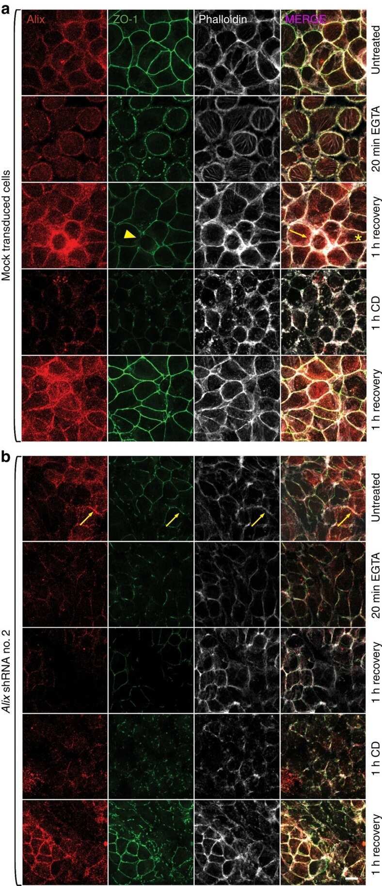
Alix-mediated assembly of the actomyosin-tight junction polarity complex preserves epithelial polarity and epithelial barrier.
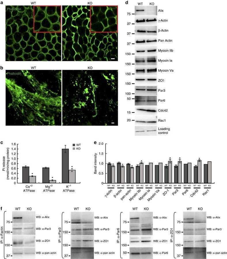
Campos Y, Qiu X, Gomero E, Wakefield R, Horner L, Brutkowski W, Han YG, Solecki D, Frase S, Bongiovanni A, d'Azzo A
Nature communications 2016 Jun 23;7:11876
Nature communications 2016 Jun 23;7:11876
The retinal phenotype of Grk1-/- is compromised by a Crb1 rd8 mutation.
Pak JS, Lee EJ, Craft CM
Molecular vision 2015;21:1281-94
Molecular vision 2015;21:1281-94
EB1 regulates tubulin and actin cytoskeletal networks at the sertoli cell blood-testis barrier in male rats: an in vitro study.
Tang EI, Mok KW, Lee WM, Cheng CY
Endocrinology 2015 Feb;156(2):680-93
Endocrinology 2015 Feb;156(2):680-93
Perfluorooctanesulfonate (PFOS) perturbs male rat Sertoli cell blood-testis barrier function by affecting F-actin organization via p-FAK-Tyr(407): an in vitro study.
Wan HT, Mruk DD, Wong CK, Cheng CY
Endocrinology 2014 Jan;155(1):249-62
Endocrinology 2014 Jan;155(1):249-62
A multiplex high-throughput gene expression assay to simultaneously detect disease and functional markers in induced pluripotent stem cell-derived retinal pigment epithelium.
Ferrer M, Corneo B, Davis J, Wan Q, Miyagishima KJ, King R, Maminishkis A, Marugan J, Sharma R, Shure M, Temple S, Miller S, Bharti K
Stem cells translational medicine 2014 Aug;3(8):911-22
Stem cells translational medicine 2014 Aug;3(8):911-22
Mesoderm is required for coordinated cell movements within zebrafish neural plate in vivo.
Araya C, Tawk M, Girdler GC, Costa M, Carmona-Fontaine C, Clarke JD
Neural development 2014 Apr 23;9:9
Neural development 2014 Apr 23;9:9
Rai14 (retinoic acid induced protein 14) is involved in regulating f-actin dynamics at the ectoplasmic specialization in the rat testis*.
Qian X, Mruk DD, Cheng CY
PloS one 2013;8(4):e60656
PloS one 2013;8(4):e60656
A novel human endogenous retroviral protein inhibits cell-cell fusion.
Sugimoto J, Sugimoto M, Bernstein H, Jinno Y, Schust D
Scientific reports 2013;3:1462
Scientific reports 2013;3:1462
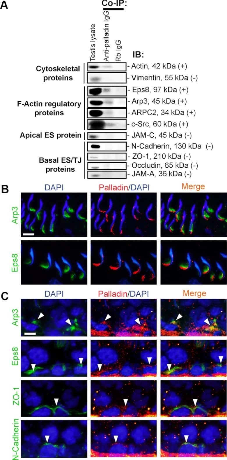
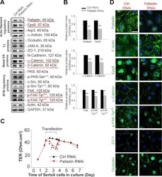
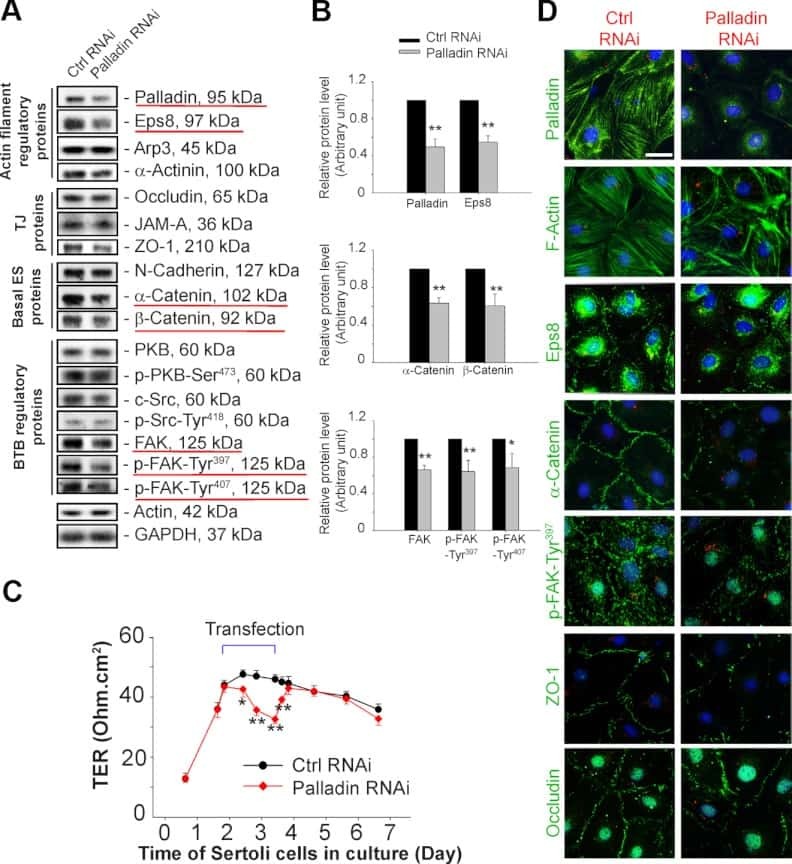
Palladin is a regulator of actin filament bundles at the ectoplasmic specialization in adult rat testes.
Qian X, Mruk DD, Wong EW, Lie PP, Cheng CY
Endocrinology 2013 May;154(5):1907-20
Endocrinology 2013 May;154(5):1907-20
Tumor necrosis factor α-mediated restructuring of the Sertoli cell barrier in vitro involves matrix metalloprotease 9 (MMP9), membrane-bound intercellular adhesion molecule-1 (ICAM-1) and the actin cytoskeleton.
Lydka M, Bilinska B, Cheng CY, Mruk DD
Spermatogenesis 2012 Oct 1;2(4):294-303
Spermatogenesis 2012 Oct 1;2(4):294-303
A mouse model for interstitial cystitis/painful bladder syndrome based on APF inhibition of bladder epithelial repair: a pilot study.
Keay S, Leitzell S, Ochrzcin A, Clements G, Zhan M, Johnson D
BMC urology 2012 Jun 8;12:17
BMC urology 2012 Jun 8;12:17
Hailey-Hailey disease and tight junctions: Claudins 1 and 4 are regulated by ATP2C1 gene encoding Ca(2+) /Mn(2+) ATPase SPCA1 in cultured keratinocytes.
Raiko L, Siljamäki E, Mahoney MG, Putaala H, Suominen E, Peltonen J, Peltonen S
Experimental dermatology 2012 Aug;21(8):586-91
Experimental dermatology 2012 Aug;21(8):586-91
Midbody accumulation through evasion of autophagy contributes to cellular reprogramming and tumorigenicity.
Kuo TC, Chen CT, Baron D, Onder TT, Loewer S, Almeida S, Weismann CM, Xu P, Houghton JM, Gao FB, Daley GQ, Doxsey S
Nature cell biology 2011 Sep 11;13(10):1214-23
Nature cell biology 2011 Sep 11;13(10):1214-23
Protective effects of nonionic triblock copolymers on bile acid-mediated epithelial barrier disruption.
Edelstein A, Fink D, Musch M, Valuckaite V, Zaborina O, Grubjesic S, Firestone MA, Matthews JB, Alverdy JC
Shock (Augusta, Ga.) 2011 Nov;36(5):451-7
Shock (Augusta, Ga.) 2011 Nov;36(5):451-7
Vascular endothelial cells cultured from patients with cerebral or uncomplicated malaria exhibit differential reactivity to TNF.
Wassmer SC, Moxon CA, Taylor T, Grau GE, Molyneux ME, Craig AG
Cellular microbiology 2011 Feb;13(2):198-209
Cellular microbiology 2011 Feb;13(2):198-209
Interleukin-1alpha is a regulator of the blood-testis barrier.
Lie PP, Cheng CY, Mruk DD
FASEB journal : official publication of the Federation of American Societies for Experimental Biology 2011 Apr;25(4):1244-53
FASEB journal : official publication of the Federation of American Societies for Experimental Biology 2011 Apr;25(4):1244-53
Adjudin-mediated Sertoli-germ cell junction disassembly affects Sertoli cell barrier function in vitro and in vivo.
Su L, Cheng CY, Mruk DD
The international journal of biochemistry & cell biology 2010 Nov;42(11):1864-75
The international journal of biochemistry & cell biology 2010 Nov;42(11):1864-75
Multipotent mesenchymal stem cells from human placenta: critical parameters for isolation and maintenance of stemness after isolation.
Semenov OV, Koestenbauer S, Riegel M, Zech N, Zimmermann R, Zisch AH, Malek A
American journal of obstetrics and gynecology 2010 Feb;202(2):193.e1-193.e13
American journal of obstetrics and gynecology 2010 Feb;202(2):193.e1-193.e13
Effects of di(2-ethylhexyl) phthalate on gap and tight junction protein expression in the testis of prepubertal rats.
Sobarzo CM, Lustig L, Ponzio R, Suescun MO, Denduchis B
Microscopy research and technique 2009 Nov;72(11):868-77
Microscopy research and technique 2009 Nov;72(11):868-77
Interleukin 1 alpha (IL1A) is a novel regulator of the blood-testis barrier in the rat.
Sarkar O, Mathur PP, Cheng CY, Mruk DD
Biology of reproduction 2008 Mar;78(3):445-54
Biology of reproduction 2008 Mar;78(3):445-54
Localization of semaphorin 3A in the rat cornea.
Morishige N, Ko JA, Liu Y, Chikama T, Nishida T
Experimental eye research 2008 Apr;86(4):669-74
Experimental eye research 2008 Apr;86(4):669-74
Effect of low fluence diode laser irradiation on the hydraulic conductivity of perfused trabecular meshwork endothelial cell monolayers.
Roberts CJ, Rivera BK, Grzybowski DM, Mahmoud AM, Weber PA
Current eye research 2007 Jul-Aug;32(7-8):625-38
Current eye research 2007 Jul-Aug;32(7-8):625-38
Dynamin II interacts with the cadherin- and occludin-based protein complexes at the blood-testis barrier in adult rat testes.
Lie PP, Xia W, Wang CQ, Mruk DD, Yan HH, Wong CH, Lee WM, Cheng CY
The Journal of endocrinology 2006 Dec;191(3):571-86
The Journal of endocrinology 2006 Dec;191(3):571-86
Disruption of Sertoli-germ cell adhesion function in the seminiferous epithelium of the rat testis can be limited to adherens junctions without affecting the blood-testis barrier integrity: an in vivo study using an androgen suppression model.
Xia W, Wong CH, Lee NP, Lee WM, Cheng CY
Journal of cellular physiology 2005 Oct;205(1):141-57
Journal of cellular physiology 2005 Oct;205(1):141-57
Apical localization of a functional TRPC3/TRPC6-Ca2+-signaling complex in polarized epithelial cells. Role in apical Ca2+ influx.
Bandyopadhyay BC, Swaim WD, Liu X, Redman RS, Patterson RL, Ambudkar IS
The Journal of biological chemistry 2005 Apr 1;280(13):12908-16
The Journal of biological chemistry 2005 Apr 1;280(13):12908-16
Adhering junction dynamics in the testis are regulated by an interplay of beta 1-integrin and focal adhesion complex-associated proteins.
Siu MK, Mruk DD, Lee WM, Cheng CY
Endocrinology 2003 May;144(5):2141-63
Endocrinology 2003 May;144(5):2141-63
No comments: Submit comment
Supportive validation
- Submitted by
- Invitrogen Antibodies (provider)
- Main image
- Experimental details
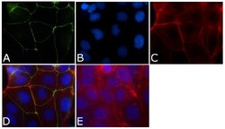
- Immunofluorescence analysis of ZO-1/TJP1 Antibody, FITC conjugate (ZO1-1A12) was done on 90% confluent log phase CaCo2 cells. The cells were fixed with 4% paraformaldehyde for 15 minutes, permeabilized with 0.25% Triton™ X-100 for 10 minutes, and blocked with 5% BSA for 1 hour at room temperature. The cells were labeled with ZO-1/TJP1 Antibody, FITC conjugate (ZO1-1A12) (Product # 33-9111) at 1µg/mL in 1% BSA and incubated for 3 hours at room temperature (Panel a: green). Nuclei (Panel b: blue) were stained with SlowFade® Gold Antifade Mountant with DAPI (Product # S36938). F-actin (Panel c: red) was stained with Alexa Fluor 594 Phalloidin (Product # A12381). Panel d is a merged image showing cell junctional localization. Panel e is a no primary antibody control. The images were captured at 40X magnification.
- Conjugate
- Green dye

Supportive validation
- Submitted by
- Invitrogen Antibodies (provider)
- Main image
- Experimental details
- NULL
- Conjugate
- Green dye

- Submitted by
- Invitrogen Antibodies (provider)
- Main image
- Experimental details
- NULL
- Conjugate
- Green dye
- Submitted by
- Invitrogen Antibodies (provider)
- Main image
- Experimental details
- NULL
- Conjugate
- Green dye
- Submitted by
- Invitrogen Antibodies (provider)
- Main image
- Experimental details
- NULL
- Conjugate
- Green dye
- Submitted by
- Invitrogen Antibodies (provider)
- Main image
- Experimental details
- NULL
- Conjugate
- Green dye
- Submitted by
- Invitrogen Antibodies (provider)
- Main image
- Experimental details
- NULL
- Conjugate
- Green dye

- Submitted by
- Invitrogen Antibodies (provider)
- Main image
- Experimental details
- NULL
- Conjugate
- Green dye

- Submitted by
- Invitrogen Antibodies (provider)
- Main image
- Experimental details
- NULL
- Conjugate
- Green dye

- Submitted by
- Invitrogen Antibodies (provider)
- Main image

- Experimental details
- NULL
- Conjugate
- Green dye
- Submitted by
- Invitrogen Antibodies (provider)
- Main image
- Experimental details
- NULL
- Conjugate
- Green dye
- Submitted by
- Invitrogen Antibodies (provider)
- Main image

- Experimental details
- NULL
- Conjugate
- Green dye
- Submitted by
- Invitrogen Antibodies (provider)
- Main image
- Experimental details
- NULL
- Conjugate
- Green dye
- Submitted by
- Invitrogen Antibodies (provider)
- Main image

- Experimental details
- NULL
- Conjugate
- Green dye
- Submitted by
- Invitrogen Antibodies (provider)
- Main image
- Experimental details
- NULL
- Conjugate
- Green dye

- Submitted by
- Invitrogen Antibodies (provider)
- Main image

- Experimental details
- NULL
- Conjugate
- Green dye
- Submitted by
- Invitrogen Antibodies (provider)
- Main image
- Experimental details
- NULL
- Conjugate
- Green dye
- Submitted by
- Invitrogen Antibodies (provider)
- Main image
- Experimental details
- NULL
- Conjugate
- Green dye
- Submitted by
- Invitrogen Antibodies (provider)
- Main image
- Experimental details
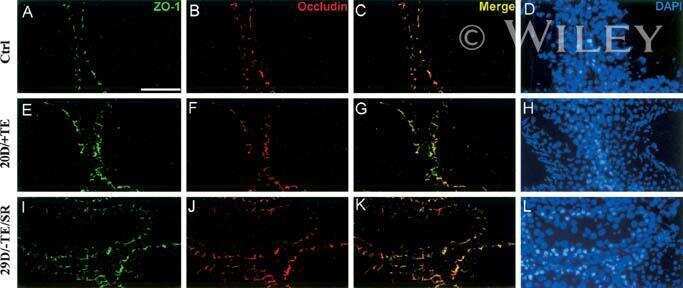
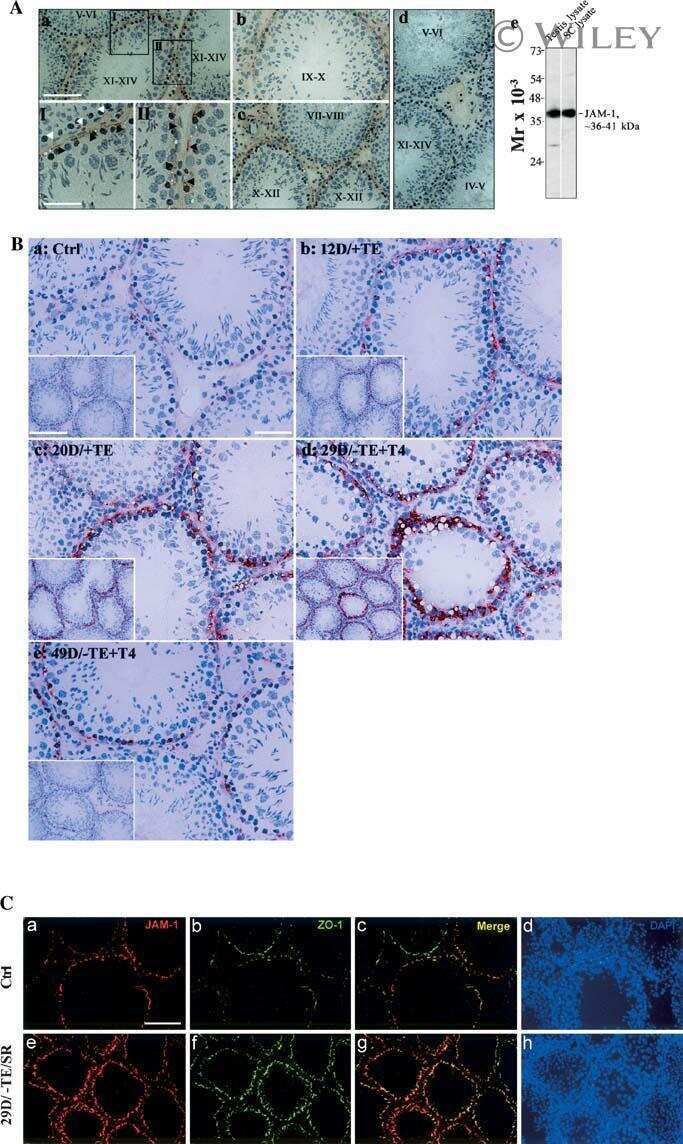
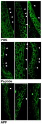
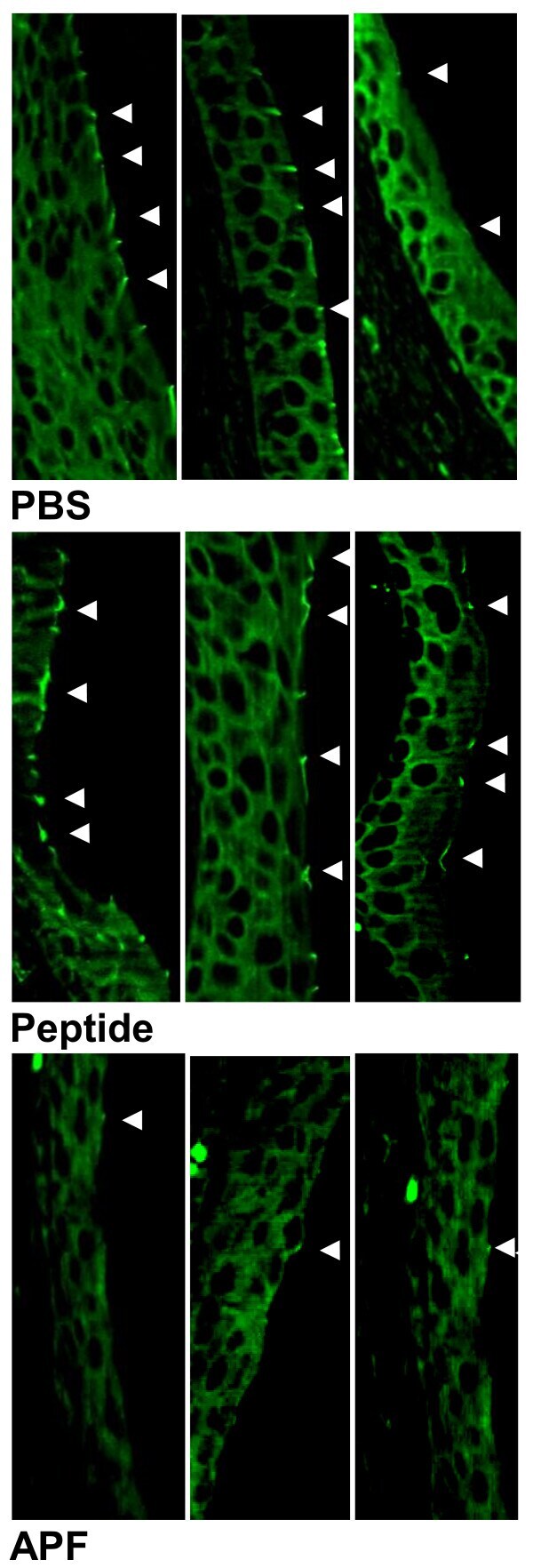
- Figure 4 Inhibition of zonula occludens type 1 (ZO-1) expression in CBA/J/Hsd mouse bladders following 14 days of treatment with as -APF. Bladder sections from mice treated with as -APF following bladder epithelial injury show decreased ZO-1 immunofluorescence staining (shown by small junctions between cells indicated by arrows) as compared to mice treated with PBS or inactive control nonglycosylated peptide. Representative data shown for the 3 mice in each treatment group from one experiment; experiment performed three times. (500X final magnification).
- Conjugate
- Green dye
- Submitted by
- Invitrogen Antibodies (provider)
- Main image
- Experimental details
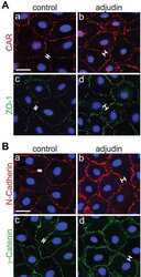
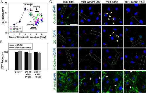
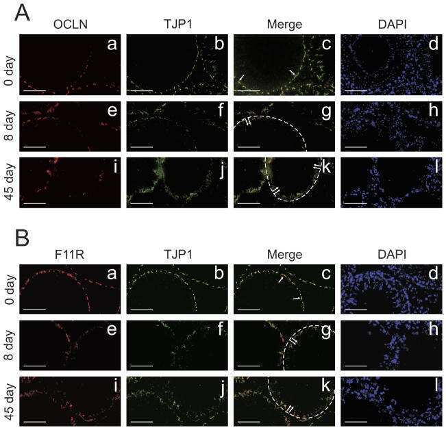
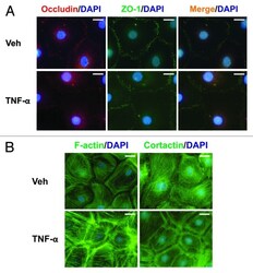
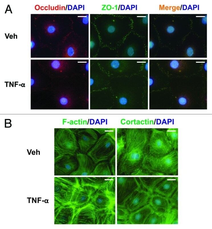
- Figure 5. Effects of TNFalpha on protein distribution within Sertoli cells. Sertoli cells (0.04 x 10 6 cells/cm 2 ) were cultured on Matrigel(tm)-coated micro cover glasses for 4 d and treated with TNFalpha (25 ng/ml) for 24 h as described in Materials and Methods. Sertoli cells were then dual-labeled for occludin (red)/ZO-1 (green), or labeled for F-actin or cortactin (both green). Corresponding images were merged to show areas of co-localization (orange, A ). Nuclei were visualized with DAPI (blue, A and B ). Scale bars, 20 mum.
- Conjugate
- Green dye
- Submitted by
- Invitrogen Antibodies (provider)
- Main image
- Experimental details
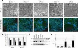
- Figure 2 Fb1 knock-down increases cell fusion in syn1 expressing cells. Fb1 -specific mRNA knock-down in BeWo choriocarcinoma cells using distinct siRNA (siFb1a and siFb1b) or their respective scrambled siRNA controls. Fb1 knock down induced a significant increase in cell fusion (a, d). (a) ZO-1-FITC and Hoechst 33342 immunocytochemistry was performed 72 hours after siRNA exposure and images are depicted in the lower row. The upper row shows matched, phase-contrast images. Scale bar represents 100 mum. Quantitative RT-PCR, immunoprecipitation and immunoblotting confirm decreases in (b) Fb1 mRNA (50-60% knock-down) and (c) protein (cell-associated and secreted) in siRNA treated Bewo cells. Expression (b) is normalized to unexposed samples cultured for similar time periods (48 or 72H). (d) Mean fusion indices for siRNA-exposed and control cells (error bars represent standard deviations; n = 4 x 5 fields). Cell fusion was assessed using phase contrast microscopy and quantitated using cell fusion indices. All observations were performed at a final magnification of 200x and total nuclei were counted per field using Leica MetaMorph image analyzing software. The number of fused syncytial aggregates and the number of nuclei in each aggregate was counted manually and fusion indices were defined as [(N-S)/T] x 100. N is the number of nuclei in syncytia, S is the number of syncytia, and T is the total number of nuclei counted. The fusion index quantitates the percentage of fusion events
- Conjugate
- Green dye

- Submitted by
- Invitrogen Antibodies (provider)
- Main image
- Experimental details
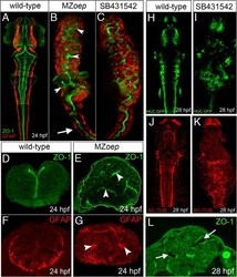
- Figure 1 MZ oep mutants have aberrant neural tube organization. (A) Projection of confocal z-series showing dorsal view of brain and anterior spinal cord. The midline ventricle is lined by ZO-1 expression (green) and the basal regions of the neuroepithelium are lined by the GFAP expression (red). (B) Projection of confocal z-series showing dorsal view of brain and anterior spinal cord from MZ oep mutant. Both apical ZO-1 and basal GFAP expression show extensive disruption to neural tube morphology in brain regions (arrowheads) but appear relatively normal in anterior spinal cord (arrow). (C) Projection of confocal z-series showing dorsal view of brain and anterior spinal cord from embryo treated with the Nodal inhibitor SB-431542. (D,E) Transverse sections show the normal single midline domain of ZO-1 appears discontinuous and more randomly oriented in MZ oep embryo. (F,G) The basal marker GFAP is expressed in ectopic foci deep from the surface of the MZ oep neural primordium. (H,I,J,K) Neurons labeled with tg(HUC-GFP) (green) and their axons labeled with Ac-tub (red) are present but disorganized in the Nodal-defective embryo brains. (L) By 28 hpf ectopic ventricles (arrowed) have opened up in the MZ oep brains. Ac-tub, anti-acetylated tubulin antibody; GFAP, glial fibrillary acidic protein; hpf, hours post fertilization; MZ oep , maternal-zygotic one-eyed pinhead ; ZO-1, zonula occludens 1.
- Conjugate
- Green dye
- Submitted by
- Invitrogen Antibodies (provider)
- Main image
- Experimental details
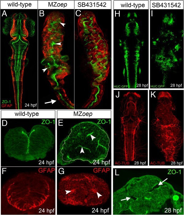
- Figure 2 Ectopic divisions do not generate the abnormal neural tube in MZ oep embryos. (A) Location and orientation of divisions monitored over a 2-hour time-lapse period of neural rod development in wild-type embryo by analyzing the expression of apical marker Pard3-GFP (green) and H2B-RFP mRNA to label nuclei (not included in image for clarity, red dumbbells indicate location and orientation of dividing cells). Wild-type neural progenitor divisions are strongly orientated along the mediolateral axis of the developing neural tube. Yellow dots outline the rod. (B) Location and orientation of divisions monitored over a 2-hour time-lapse period of neural rod development in MZ oep embryo. (C,D) Orientation plots of divisions in wild-type and MZ oep embryos. (E,F,G) Dorsal projections of ventricle morphology (ZO-1, green) at 24 hpf in wild-type, MZ oep embryos and MZ oep embryos treated with CDIs (hydroxyurea and aphidicolin). Blocking division does not rescue ventricle morphology. PH3 staining of mitotic figures (purple) used to calculate efficiency of division block. (H) Transverse section of brain from MZ oep embryo treated with division blockers. (I) Quantification of divisions in wild-type, MZ oep and MZ oep division blocked embryos. Number of cell divisions between wild-type 194 and MZ oep 206, P = 0.5129 Student's t -test and between MZ oep 206 and MZ oep + CDI 39, *** P
- Conjugate
- Green dye

- Submitted by
- Invitrogen Antibodies (provider)
- Main image
- Experimental details
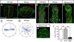
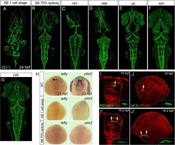
- Figure 4 Loss of mesoderm, but not Nodal signaling, leads to neural tube defects. (A,B,C,D,E,F,G) Projection of confocal z-series showing dorsal view of 24 hpf zebrafish embryos staining for the apical marker ZO-1 (green). (A) Disrupted ventricular organization of embryo treated with the Nodal inhibitor SB-431542 from the one-cell stage. (B) Treatment with SB-431542 from 70% epiboly does not cause neural tube defects. (C,D) Ventricle organization in Nodal mutants cyc and oep is largely normal. (E,F) Ventricle organization in ntl and syu mutants is normal. (G) Ventricle organization in the endoderm mutant cas mutants is normal. (H) Expression of the Nodal-dependent markers lefty and pitx2 show the SB-431542 drug is an efficient blocker of Nodal signaling. All embryos between 21 and 23 hpf, dorsal view. Arrowhead in (H) indicates asymmetric lefty expression at 21 hpf. (I,I') Two frames from time-lapse sequence show transplanted MZ oep cells (green) integrate and divide to make mirror-image daughters just like host cells in a wild-type embryo at 15 hpf. (J,J') Two frames at 15 hpf from time-lapse sequence show wild-type cells (green) divide to make mirror-image daughters after transplantation to a MZ oep embryo. Unlike divisions in wild-type embryos, however, these divisions can be far from their normal location at the midline. hpf, hours post fertilization; MZ oep , maternal-zygotic one-eyed pinhead ; wt, wild-type; ZO-1, zonula occludens 1.
- Conjugate
- Green dye
- Submitted by
- Invitrogen Antibodies (provider)
- Main image
- Experimental details
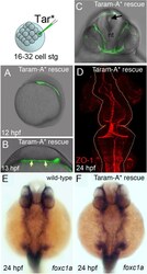
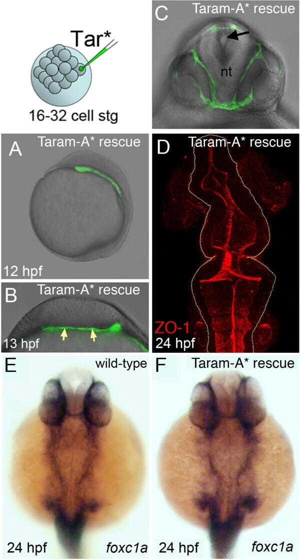
- Figure 5 Mesoderm rescues neural tube morphogenesis in MZ oep embryos. (A) Lateral view of an MZ oep embryo previously injected with the activated form of the TGF-beta receptor Taram-A* (Tar*) into a single blastomere. Rescued head mesoderm expresses GFP (green). (B) Transverse section of Taram-A*-injected embryos show rescued mesoderm (green and arrowed) underlying neural plate at 13 hpf. (C) By 24 hpf rescued mesoderm almost surrounds the neural tube (nt) in MZ oep embryos. The morphology of the neural tube is rescued and contains a single well-defined midline lumen (black arrow). (D) Rescued neural lumen morphology revealed by ZO-1 expression. (E,F) Identity, distribution and presence of rescued mesoderm, confirmed by the mesendoderm marker foxc1a , in wild-type and Taram-A*-injected MZ oep embryos. GFP, green fluorescent protein; hpf, hours post fertilization; MZ oep , maternal-zygotic one-eyed pinhead ; TGF-beta, transforming growth factor beta; ZO-1, zonula occludens 1.
- Conjugate
- Green dye
- Submitted by
- Invitrogen Antibodies (provider)
- Main image
- Experimental details
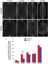
- Figure 9 Apical polarity development in MZ oep . ZO-1 staining in (A,B,C,D) wild-type and (E,F,G,H) MZ oep embryos at different stages of neurulation. All images are maximum confocal projections of six consecutive z-levels taken from a dorsal view at posterior hindbrain and anterior spinal cord regions. Anterior is up. White arrows indicate the dorsal midline of the embryo. The ZO-1 staining surrounding the edge of the neural tissue is from the polarized enveloping layer overlying the neural tissue. Scale bar is 50 mum. (I) The average number of pixels of ZO-1 staining over time for MZ oep and wild-type embryos. At 13.5 hpf the number of ZO-1 puncta in MZ oep embryos is significantly different to wild-type (* P
- Conjugate
- Green dye

- Submitted by
- Invitrogen Antibodies (provider)
- Main image
- Experimental details
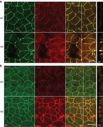
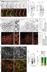
- Fig. 1 Defects in junctional remodelling upon inactivation of PI3Kalpha in endothelial cells. a Lateral views of intersomitic vessels (ISV) in vehicle and GDC-0326 (50 muM)-treated transgenic Tg( kdrl:EGFP ) s843 (shown in red) embryos stained for ZO-1 (green) at 33 h post fertilization (hpf). Single ZO-1 staining is shown in upper row. DA refers to dorsal aorta and DLAV refers to dorsal longitudinal anastomotic vessels. White lines indicate ZO-1 negative staining; punctuate white lines indicate elongation of junction; yellow arrowheads show ring-shape junctions. b Quantification of the length of the dorsal part of the ISVs without ZO-1 (left graph) and length of the ISVs with continuous ZO-1 staining (right graph) in vehicle and GDC-0326 treated embryos ( n >= 54 ISVs per treatment). c Representative maximum intensity projections of anti-VE-cadherin (green) and isolectin B4 (IB4, red) immunostained control and Pik3ca KD/iDeltaEC mouse retinas at P7. Single channel is shown in upper row. Yellow islets show higher magnification of selected regions shown to the right. Yellow arrowheads indicate vascular segments without VE-cadherin staining; red asterisks indicate VE-cadherin-positive isolated rings or single-dots within the vascular tubes, indicating cell-cell junctional contacts that have not elongated. d , e Quantitative number of VE-cadherin-negative vessels (junctional gaps) per unit area ( n >= 5 retinas per genotype) ( d ) and length of vessel structures wi
- Conjugate
- Green dye
- Submitted by
- Invitrogen Antibodies (provider)
- Main image
- Experimental details
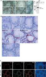
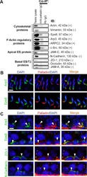
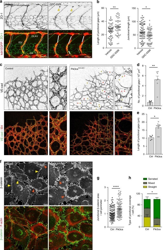
- Figure 3 Rai14 is an apical and basal ES protein in the rat testis. Dual-labeled immunofluorescence analysis was used to assess co-localization of Rai14 (red) with constituent protein laminin-gamma3 chain (green), actin regulatory protein Arp3 (green), actin binding protein drebrin E (green) and actin filament cross-linking protein palladin (green) at the apical ES in stage VII tubules when these proteins were all highly expressed. It was found that Rai14 indeed partially co-localized with each of these apical ES proteins (see ""orange yellow"" in merged images) as shown in ( A ). Rai14 (red) also partially co-localized with basal ES/TJ protein ZO-1 (see ""yellow"" arrowheads), and to a lesser extent with N-cadherin (see ""white"" arrowheads) in stage IV-V tubules when these proteins were highly expressed as shown in ( B ). Bar = 10 um in ( A ) or ( B ), which applies all other images in ( A ) and ( B ).
- Conjugate
- Green dye

- Submitted by
- Invitrogen Antibodies (provider)
- Main image

- Experimental details
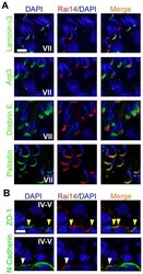
- Immunostaining of Cells in the Rocked Device, Multi-cell line Static Function Control, and Single-cell line Static Function Controls. Fluorescent confocal images of the nonbarrier cell lines (A549 and Caco2) were observed after immunostaining for surfactant (red) and tight junctions (protein ZO-1, green). The nuclei were counter-stained (DAPI, blue). The cultures were observed under a water immersion 25x collared objective and further magnified with the Zen program. Scale bars are present on the micrographs.
- Conjugate
- Green dye
- Submitted by
- Invitrogen Antibodies (provider)
- Main image
- Experimental details
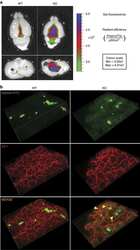
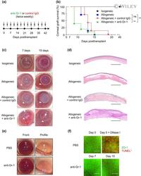
- 1 FIGURE Depletion of MDSC accelerates corneal allograft rejection. (A) Graphic illustration of experimental design. Isogeneic (BALB/c to BALB/c) and allogeneic (C57BL/6 to BALB/c) corneal transplantation was performed on day 0. Anti-Gr-1 antibody or control IgG was interperitoneally administered twice weekly for 6 weeks after transplantation. (B) Kaplan-Meier survival curves of isografts ( n = 5) and allografts ( n = 10 each group). **, P < .01; ns, not significant; log-rank tests. (C) Representative slim-lamp photographs (front view) of graft-bearing eyeballs on day 7 and day 15 posttransplant. Dashed circles outline corneal grafts. White arrows and arrowheads denote representative neovessels in the corneal graft and the graft bed, respectively. Open arrowheads in photographs of the isograft denote representative blood vessels in the iris. (D) H&E stain of corneal grafts on day 15 posttransplant from mice treated as in (A-C). Scale bars, 200 mum. (E) Representative slim-lamp photographs (front view and profile view) of normal (graft-free) eyeballs from mice treated with anti-Gr-1 antibody or PBS control on day 15 posttransplant as in (C). (F) Representative immunohistochemistry staining of the tight junction protein zonula occludens-1 (ZO-1) and TUNEL staining of corneal endothelium of normal eyeballs from untreated mice or mice treated with anti-Gr-1 antibody on day 7 and day 15 posttransplant as in (A-C). Nuclei of apoptotic cells were stained red in TUNEL assay and DNase
- Conjugate
- Green dye

- Submitted by
- Invitrogen Antibodies (provider)
- Main image
- Experimental details
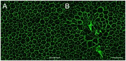
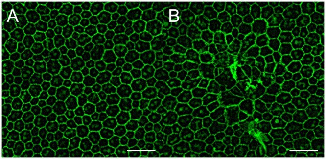
- 10.1371/journal.pone.0246115.g006 Fig 6 Alterations of the retinal pigment epithelium after suturing of the vortex veins. (A) Immunohistochemistry using anti-ZO-1 antibody for RPE flatmount in the control C57BL/6 mouse eye shows well organized and packed RPE cells. (B) 7 days after the vortex vein suturing, focal RPE cell degeneration is observed. Scale bar: 50mum.
- Conjugate
- Green dye
- Submitted by
- Invitrogen Antibodies (provider)
- Main image

- Experimental details
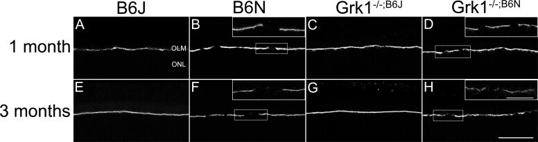
- Figure 6 Immunohistochemistry analysis of retinal ZO-1. Primary antibody to ZO-1 (1:1,000) followed by secondary antibody Alexa Fluor donkey anti-rabbit 488 (1:500) identifies immunohistochemical staining in mouse retina sections at 1 month ( A - D ) and 3 months ( E - H ) localized to the OLM. A, C, E, G : The OLM in the B6J background retinas is unbroken. B, D, F, H : The OLM in the B6N background retinas is discontinuous. The top-right corners show higher magnification images. Lower magnification images were taken with a 20X objective lens (scale bar, 50 um); higher magnification images were taken at 100X objective (scale bar, 20 um). OLM=outer limiting membrane; ONL=outer nuclear layer.
- Conjugate
- Green dye