 Explore
 Validate
 Learn
Learn Western blot
 Immunocytochemistry
Antibody data
- Antibody Data
- Antigen structure
- References [9]
- Comments [0]
- Validations
- Western blot [1]
- Immunocytochemistry [1]
- Immunohistochemistry [1]
Submit
Validation data
Reference
Comment
Report error
- Product number
- HPA001636 - Provider product page

- Provider
- Atlas Antibodies
- Proper citation
- Atlas Antibodies Cat#HPA001636, RRID:AB_1080762
- Product name
- Anti-TJP1
- Antibody type
- Polyclonal
- Description
- Polyclonal Antibody against Human TJP1, Gene description: tight junction protein 1, Alternative Gene Names: DKFZp686M05161, MGC133289, ZO-1, Validated applications: ICC, IHC, WB, Uniprot ID: Q07157, Storage: Store at +4°C for short term storage. Long time storage is recommended at -20°C.
- Reactivity
- Human
- Host
- Rabbit
- Conjugate
- Unconjugated
- Isotype
- IgG
- Vial size
- 100 µl
- Concentration
- 0.1 mg/ml
- Storage
- Store at +4°C for short term storage. Long time storage is recommended at -20°C.
- Handling
- The antibody solution should be gently mixed before use.
Submitted references Triangular Silver Nanoplates as a Bioanalytical Tool: Potential COVID-19 Detection.
Moderate Dose Irradiation Induces DNA Damage and Impairments of Barrier and Host Defense in Nasal Epithelial Cells in vitro
High expression of tight junction protein 1 as a predictive biomarker for bladder cancer grade and staging
Host Antiviral Response Suppresses Ciliogenesis and Motile Ciliary Functions in the Nasal Epithelium
Toll‐like receptor 2 stimulation augments esophageal barrier integrity
Disruption of gut integrity and permeability contributes to enteritis in a fish-parasite model: a story told from serum metabolomics
Induced cortical tension restores functional junctions in adhesion-defective carcinoma cells
A recellularized human colon model identifies cancer driver genes
Expansive Generation of Functional Airway Epithelium From Human Embryonic Stem Cells
Rodriguez Barroso LG, Lanzagorta Garcia E, Mojicevic M, Alkan Tas B, Huerta M, Pogue R, Devine DM, Brennan-Fournet M
International journal of molecular sciences 2023 Jul 26;24(15)
International journal of molecular sciences 2023 Jul 26;24(15)
Moderate Dose Irradiation Induces DNA Damage and Impairments of Barrier and Host Defense in Nasal Epithelial Cells in vitro
Yang Y, Liu J, Liu Y, Ong H, Chen Q, Chen C, Thong M, Xu X, Zhou S, Qiu Q, Wang D
Journal of Inflammation Research 2022;Volume 15
Journal of Inflammation Research 2022;Volume 15
High expression of tight junction protein 1 as a predictive biomarker for bladder cancer grade and staging
Lee Y, Tsai K, Liao J, Kuo W, Chang Y, Yang Y
Scientific Reports 2022;12(1)
Scientific Reports 2022;12(1)
Host Antiviral Response Suppresses Ciliogenesis and Motile Ciliary Functions in the Nasal Epithelium
Chen Q, Tan K, Liu J, Ong H, Zhou S, Huang H, Chen H, Ong Y, Thong M, Chow V, Qiu Q, Wang D
Frontiers in Cell and Developmental Biology 2020;8
Frontiers in Cell and Developmental Biology 2020;8
Toll‐like receptor 2 stimulation augments esophageal barrier integrity
Ruffner M, Song L, Maurer K, Shi L, Carroll M, Wang J, Muir A, Spergel J, Sullivan K
Allergy 2019;74(12):2449-2460
Allergy 2019;74(12):2449-2460
Disruption of gut integrity and permeability contributes to enteritis in a fish-parasite model: a story told from serum metabolomics
Sitjà-Bobadilla A, Gil-Solsona R, Estensoro I, Piazzon M, Martos-Sitcha J, Picard-Sánchez A, Fuentes J, Sancho J, Calduch-Giner J, Hernández F, Pérez-Sánchez J
Parasites & Vectors 2019;12(1)
Parasites & Vectors 2019;12(1)
Induced cortical tension restores functional junctions in adhesion-defective carcinoma cells
Ito S, Okuda S, Abe M, Fujimoto M, Onuki T, Nishimura T, Takeichi M
Nature Communications 2017;8(1)
Nature Communications 2017;8(1)
A recellularized human colon model identifies cancer driver genes
Chen H, Wei Z, Sun J, Bhattacharya A, Savage D, Serda R, Mackeyev Y, Curley S, Bu P, Wang L, Chen S, Cohen-Gould L, Huang E, Shen X, Lipkin S, Copeland N, Jenkins N, Shuler M
Nature Biotechnology 2016;34(8):845-851
Nature Biotechnology 2016;34(8):845-851
Expansive Generation of Functional Airway Epithelium From Human Embryonic Stem Cells
McIntyre B, Alev C, Mechael R, Salci K, Lee J, Fiebig-Comyn A, Guezguez B, Wu Y, Sheng G, Bhatia M
Stem Cells Translational Medicine 2014;3(1):7-17
Stem Cells Translational Medicine 2014;3(1):7-17
No comments: Submit comment
Enhanced validation
- Submitted by
- Atlas Antibodies (provider)
- Enhanced method
- Recombinant expression validation
- Main image
- Experimental details
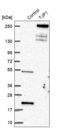
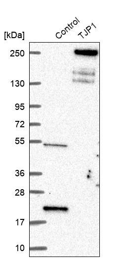
- Western blot analysis in control (vector only transfected HEK293T lysate) and TJP1 over-expression lysate (Co-expressed with a C-terminal myc-DDK tag (~3.1 kDa) in mammalian HEK293T cells, LY418804).
- Sample type
- Human
- Protocol
- Protocol
Supportive validation
- Submitted by
- Atlas Antibodies (provider)
- Main image
- Experimental details
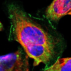
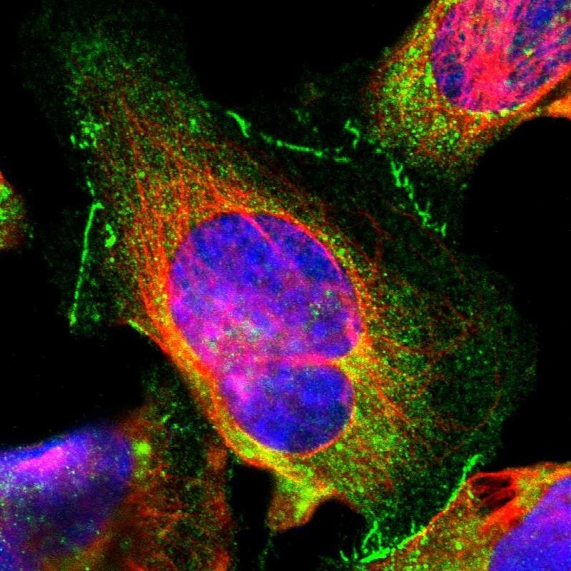
- Immunofluorescent staining of human cell line U-2 OS shows localization to cytosol & cell junctions.
- Sample type
- Human
Supportive validation
- Submitted by
- Atlas Antibodies (provider)
- Enhanced method
- Orthogonal validation
- Main image
- Experimental details
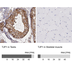
- Immunohistochemistry analysis in human testis and skeletal muscle tissues using HPA001636 antibody. Corresponding TJP1 RNA-seq data are presented for the same tissues.
- Sample type
- Human
- Protocol
- Protocol
