Explore
 Validate
 Learn
Learn Western blot
 Immunohistochemistry
Antibody data
- Antibody Data
- Antigen structure
- References [2]
- Comments [0]
- Validations
- Immunohistochemistry [1]
Submit
Validation data
Reference
Comment
Report error
- Product number
- AMAb90911 - Provider product page

- Provider
- Atlas Antibodies
- Proper citation
- Atlas Antibodies Cat#AMAb90911, RRID:AB_2665722
- Product name
- Anti-ITGAM
- Antibody type
- Monoclonal
- Description
- Monoclonal Antibody against Human ITGAM, Clone ID: CL1719, Gene description: integrin, alpha M (complement component 3 receptor 3 subunit), Alternative Gene Names: CD11B, CR3A, MAC-1, Validated applications: WB, IHC, Uniprot ID: P11215, Storage: Store at +4°C for short term storage. Long time storage is recommended at -20°C.
- Reactivity
- Human
- Host
- Mouse
- Conjugate
- Unconjugated
- Isotype
- IgG
- Antibody clone number
- CL1719
- Vial size
- 100 µl
- Concentration
- 0.1 mg/ml
- Storage
- Store at +4°C for short term storage. Long time storage is recommended at -20°C.
- Handling
- The antibody solution should be gently mixed before use.
Submitted references Exercise-induced crosstalk between immune cells and adipocytes in humans: Role of oncostatin-M.
Distinctive exercise-induced inflammatory response and exerkine induction in skeletal muscle of people with type 2 diabetes
Dollet L, Lundell LS, Chibalin AV, Pendergrast LA, Pillon NJ, Lansbury EL, Elmastas M, Frendo-Cumbo S, Jalkanen J, de Castro Barbosa T, Cervone DT, Caidahl K, Dmytriyeva O, Deshmukh AS, Barrès R, Rydén M, Wallberg-Henriksson H, Zierath JR, Krook A
Cell reports. Medicine 2024 Jan 16;5(1):101348
Cell reports. Medicine 2024 Jan 16;5(1):101348
Distinctive exercise-induced inflammatory response and exerkine induction in skeletal muscle of people with type 2 diabetes
Pillon N, Smith J, Alm P, Chibalin A, Alhusen J, Arner E, Carninci P, Fritz T, Otten J, Olsson T, van Doorslaer de ten Ryen S, Deldicque L, Caidahl K, Wallberg-Henriksson H, Krook A, Zierath J
Science Advances 2022;8(36)
Science Advances 2022;8(36)
No comments: Submit comment
Supportive validation
- Submitted by
- Atlas Antibodies (provider)
- Enhanced method
- Orthogonal validation
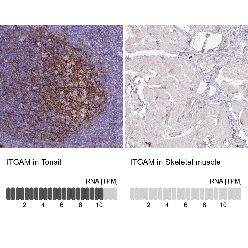
- Main image
- Experimental details
- Immunohistochemistry analysis in human tonsil and skeletal muscle tissues using AMAb90911 antibody. Corresponding ITGAM RNA-seq data are presented for the same tissues.
- Sample type
- Human
- Protocol
- Protocol