Explore
 Validate
 Learn
Learn Flow cytometry
Antibody data
- Antibody Data
- Antigen structure
- References [24]
- Comments [0]
- Validations
- Flow cytometry [2]
- Other assay [9]
Submit
Validation data
Reference
Comment
Report error
- Product number
- 11-0113-42 - Provider product page

- Provider
- Invitrogen Antibodies
- Product name
- CD11b (activation epitope) Monoclonal Antibody (CBRM1/5), FITC, eBioscience™
- Antibody type
- Monoclonal
- Antigen
- Other
- Description
- Description: The CBRM1/5 monoclonal antibody reacts with an activation-specific epitope of human Mac-1. CBRM1/5 binds a subset of Mac-1 molecules on neutrophils and monocytes after stimulation with chemoattractants or phorbol esters but does not recognize Mac-1 on resting myeloid cells. Through interactions with its ligands, Mac-1 participates in adhesive cell interactions. The epitope recognized by this mAb localizes to the I domain on the alpha chain of Mac-1 very close to the ligand binding site in a region that is widely exposed. CBRM1/5 blocks Mac-1 dependent adhesion to fibrinogen and ICAM-1 and inhibits chemoattractant-stimulated adhesion of eosinophils to the Intercellular Adhesion Molecule-1 (ICAM-1). It should be noted that low level activation may occur during processing of freshly drawn blood. Therefore the CBRM1.5 antibody may exhibit some binding to Mac-1 in these unstimulated samples. However, higher levels of Mac-1 expression are observed in activated samples when compared to unstimulated cells. Applications Reported: The CBRM1/5 antibody has been reported for use in flow cytometric analysis. Applications Tested: The CBRM1/5 antibody has been pre-titrated and tested by flow cytometric analysis of resting and activated normal human peripheral blood cells. This can be used at 5 µL (1 µg) per test. A test is defined as the amount (µg) of antibody that will stain a cell sample in a final volume of 100 µL. Cell number should be determined empirically but can range from 10^5 to 10^8 cells/test. Excitation: 488 nm; Emission: 520 nm; Laser: Blue Laser. Filtration: 0.2 µm post-manufacturing filtered.
- Reactivity
- Human
- Host
- Mouse
- Conjugate
- Green dye
- Isotype
- IgG
- Antibody clone number
- CBRM1/5
- Vial size
- 100 Tests
- Concentration
- 5 μL/Test
- Storage
- 4°C, store in dark, DO NOT FREEZE!
Submitted references Lipoxins modulate neutrophil oxidative burst, integrin expression and lymphatic transmigration differentially in human health and atherosclerosis.
Upregulation of CD3ζ and L-selectin in antigen-specific cytotoxic T lymphocytes by eliminating myeloid-derived suppressor cells with doxorubicin to improve killing efficacy of neuroblastoma cells in vitro.
RAS-Responsive Element-Binding Protein 1 Blocks the Granulocytic Differentiation of Myeloid Leukemia Cells.
Deficiency of FAM3D (Family With Sequence Similarity 3, Member D), A Novel Chemokine, Attenuates Neutrophil Recruitment and Ameliorates Abdominal Aortic Aneurysm Development.
Human decidua mesenchymal stem cells regulate decidual natural killer cell function via interactions between collagen and leukocyte‑associated immunoglobulin‑like receptor 1.
TLR2 activation induces antioxidant defence in human monocyte-macrophage cell line models.
A Member of the Nuclear Receptor Superfamily, Designated as NR2F2, Supports the Self-Renewal Capacity and Pluripotency of Human Bone Marrow-Derived Mesenchymal Stem Cells.
Histidine-Rich Glycoprotein Prevents Septic Lethality through Regulation of Immunothrombosis and Inflammation.
Differential in vivo activation of monocyte subsets during low-grade inflammation through experimental endotoxemia in humans.
A novel PAD4/SOX4/PU.1 signaling pathway is involved in the committed differentiation of acute promyelocytic leukemia cells into granulocytic cells.
NLS‑RARα modulates acute promyelocytic leukemia NB4 cell proliferation and differentiation via the PI3K/AKT pathway.
Serum biomarkers VEGF-C and IL-6 are associated with severe human Peripheral Artery Stenosis.
Pathways related to PMA-differentiated THP1 human monocytic leukemia cells revealed by RNA-Seq.
MIR125B1 represses the degradation of the PML-RARA oncoprotein by an autophagy-lysosomal pathway in acute promyelocytic leukemia.
Fluid shear stress increases neutrophil activation via platelet-activating factor.
Adiponectin inhibits neutrophil phagocytosis of Escherichia coli by inhibition of PKB and ERK 1/2 MAPK signalling and Mac-1 activation.
c-myc but not Hif-1α-dependent downregulation of VEGF influences the proliferation and differentiation of HL-60 cells induced by ATRA.
Common genetic variations in the NALP3 inflammasome are associated with delayed apoptosis of human neutrophils.
Gu-4 suppresses affinity and avidity modulation of CD11b and improves the outcome of mice with endotoxemia and sepsis.
Endogenous PMN sialidase activity exposes activation epitope on CD11b/CD18 which enhances its binding interaction with ICAM-1.
Outside-in signal transmission by conformational changes in integrin Mac-1.
CD44-mediated phagocytosis induces inside-out activation of complement receptor-3 in murine macrophages.
Antineutrophil cytoplasm antibody-stimulated neutrophil adhesion depends on diacylglycerol kinase-catalyzed phosphatidic acid formation.
ANCA induces beta2 integrin and CXC chemokine-dependent neutrophil-endothelial cell interactions that mimic those of highly cytokine-activated endothelium.
Kraft JD, Blomgran R, Bergström I, Soták M, Clark M, Rani A, Rajan MR, Dalli J, Nyström S, Quiding-Järbrink M, Bromberg J, Skoog P, Börgeson E
FASEB journal : official publication of the Federation of American Societies for Experimental Biology 2022 Mar;36(3):e22173
FASEB journal : official publication of the Federation of American Societies for Experimental Biology 2022 Mar;36(3):e22173
Upregulation of CD3ζ and L-selectin in antigen-specific cytotoxic T lymphocytes by eliminating myeloid-derived suppressor cells with doxorubicin to improve killing efficacy of neuroblastoma cells in vitro.
Xu W, Li S, Li M, Zhou H, Yang X
Journal of clinical laboratory analysis 2022 Jan;36(1):e24158
Journal of clinical laboratory analysis 2022 Jan;36(1):e24158
RAS-Responsive Element-Binding Protein 1 Blocks the Granulocytic Differentiation of Myeloid Leukemia Cells.
Yao J, Zhong L, Zhong P, Liu D, Yuan Z, Liu J, Yao S, Zhao Y, Chen M, Li L, Liu L, Liu B
Oncology research 2019 Jul 12;27(7):809-818
Oncology research 2019 Jul 12;27(7):809-818
Deficiency of FAM3D (Family With Sequence Similarity 3, Member D), A Novel Chemokine, Attenuates Neutrophil Recruitment and Ameliorates Abdominal Aortic Aneurysm Development.
He L, Fu Y, Deng J, Shen Y, Wang Y, Yu F, Xie N, Chen Z, Hong T, Peng X, Li Q, Zhou J, Han J, Wang Y, Xi J, Kong W
Arteriosclerosis, thrombosis, and vascular biology 2018 Jul;38(7):1616-1631
Arteriosclerosis, thrombosis, and vascular biology 2018 Jul;38(7):1616-1631
Human decidua mesenchymal stem cells regulate decidual natural killer cell function via interactions between collagen and leukocyte‑associated immunoglobulin‑like receptor 1.
Fu Q, Man X, Yu M, Chu Y, Luan X, Piao H, Xue J, Jin C
Molecular medicine reports 2017 Sep;16(3):2791-2798
Molecular medicine reports 2017 Sep;16(3):2791-2798
TLR2 activation induces antioxidant defence in human monocyte-macrophage cell line models.
Karwaciak I, Gorzkiewicz M, Bartosz G, Pulaski L
Oncotarget 2017 Aug 15;8(33):54243-54264
Oncotarget 2017 Aug 15;8(33):54243-54264
A Member of the Nuclear Receptor Superfamily, Designated as NR2F2, Supports the Self-Renewal Capacity and Pluripotency of Human Bone Marrow-Derived Mesenchymal Stem Cells.
Zhu N, Wang H, Wang B, Wei J, Shan W, Feng J, Huang H
Stem cells international 2016;2016:5687589
Stem cells international 2016;2016:5687589
Histidine-Rich Glycoprotein Prevents Septic Lethality through Regulation of Immunothrombosis and Inflammation.
Wake H, Mori S, Liu K, Morioka Y, Teshigawara K, Sakaguchi M, Kuroda K, Gao Y, Takahashi H, Ohtsuka A, Yoshino T, Morimatsu H, Nishibori M
EBioMedicine 2016 Jul;9:180-194
EBioMedicine 2016 Jul;9:180-194
Differential in vivo activation of monocyte subsets during low-grade inflammation through experimental endotoxemia in humans.
Thaler B, Hohensinner PJ, Krychtiuk KA, Matzneller P, Koller L, Brekalo M, Maurer G, Huber K, Zeitlinger M, Jilma B, Wojta J, Speidl WS
Scientific reports 2016 Jul 22;6:30162
Scientific reports 2016 Jul 22;6:30162
A novel PAD4/SOX4/PU.1 signaling pathway is involved in the committed differentiation of acute promyelocytic leukemia cells into granulocytic cells.
Song G, Shi L, Guo Y, Yu L, Wang L, Zhang X, Li L, Han Y, Ren X, Guo Q, Bi K, Jiang G
Oncotarget 2016 Jan 19;7(3):3144-57
Oncotarget 2016 Jan 19;7(3):3144-57
NLS‑RARα modulates acute promyelocytic leukemia NB4 cell proliferation and differentiation via the PI3K/AKT pathway.
Song H, Li L, Zhong L, Yang R, Jiang K, Yang X, Liu B
Molecular medicine reports 2016 Dec;14(6):5495-5500
Molecular medicine reports 2016 Dec;14(6):5495-5500
Serum biomarkers VEGF-C and IL-6 are associated with severe human Peripheral Artery Stenosis.
Chen J, Han L, Xu X, Tang H, Wang H, Wei B
Journal of inflammation (London, England) 2015;12:50
Journal of inflammation (London, England) 2015;12:50
Pathways related to PMA-differentiated THP1 human monocytic leukemia cells revealed by RNA-Seq.
Zeng C, Wang W, Yu X, Yang L, Chen S, Li Y
Science China. Life sciences 2015 Dec;58(12):1282-7
Science China. Life sciences 2015 Dec;58(12):1282-7
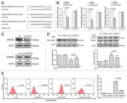
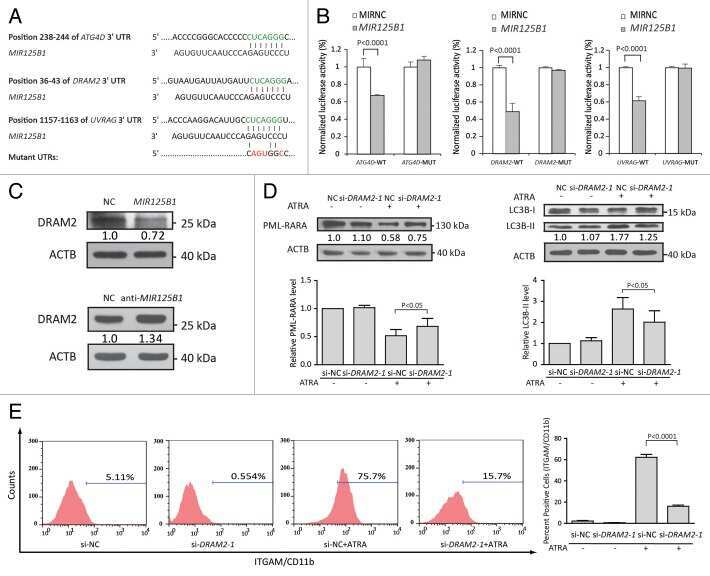
MIR125B1 represses the degradation of the PML-RARA oncoprotein by an autophagy-lysosomal pathway in acute promyelocytic leukemia.
Zeng CW, Chen ZH, Zhang XJ, Han BW, Lin KY, Li XJ, Wei PP, Zhang H, Li Y, Chen YQ
Autophagy 2014 Oct 1;10(10):1726-37
Autophagy 2014 Oct 1;10(10):1726-37
Fluid shear stress increases neutrophil activation via platelet-activating factor.
Mitchell MJ, Lin KS, King MR
Biophysical journal 2014 May 20;106(10):2243-53
Biophysical journal 2014 May 20;106(10):2243-53
Adiponectin inhibits neutrophil phagocytosis of Escherichia coli by inhibition of PKB and ERK 1/2 MAPK signalling and Mac-1 activation.
Rossi A, Lord J
PloS one 2013;8(7):e69108
PloS one 2013;8(7):e69108
c-myc but not Hif-1α-dependent downregulation of VEGF influences the proliferation and differentiation of HL-60 cells induced by ATRA.
Song G, Li Y, Zhang Z, Ren X, Li H, Zhang W, Wei R, Pan S, Shi L, Bi K, Jiang G
Oncology reports 2013 Jun;29(6):2378-84
Oncology reports 2013 Jun;29(6):2378-84
Common genetic variations in the NALP3 inflammasome are associated with delayed apoptosis of human neutrophils.
Blomgran R, Patcha Brodin V, Verma D, Bergström I, Söderkvist P, Sjöwall C, Eriksson P, Lerm M, Stendahl O, Särndahl E
PloS one 2012;7(3):e31326
PloS one 2012;7(3):e31326
Gu-4 suppresses affinity and avidity modulation of CD11b and improves the outcome of mice with endotoxemia and sepsis.
Yan T, Li Q, Zhou H, Zhao Y, Yu S, Xu G, Yin Z, Li Z, Zhao Z
PloS one 2012;7(2):e30110
PloS one 2012;7(2):e30110
Endogenous PMN sialidase activity exposes activation epitope on CD11b/CD18 which enhances its binding interaction with ICAM-1.
Feng C, Zhang L, Almulki L, Faez S, Whitford M, Hafezi-Moghadam A, Cross AS
Journal of leukocyte biology 2011 Aug;90(2):313-21
Journal of leukocyte biology 2011 Aug;90(2):313-21
Outside-in signal transmission by conformational changes in integrin Mac-1.
Lefort CT, Hyun YM, Schultz JB, Law FY, Waugh RE, Knauf PA, Kim M
Journal of immunology (Baltimore, Md. : 1950) 2009 Nov 15;183(10):6460-8
Journal of immunology (Baltimore, Md. : 1950) 2009 Nov 15;183(10):6460-8
CD44-mediated phagocytosis induces inside-out activation of complement receptor-3 in murine macrophages.
Vachon E, Martin R, Kwok V, Cherepanov V, Chow CW, Doerschuk CM, Plumb J, Grinstein S, Downey GP
Blood 2007 Dec 15;110(13):4492-502
Blood 2007 Dec 15;110(13):4492-502
Antineutrophil cytoplasm antibody-stimulated neutrophil adhesion depends on diacylglycerol kinase-catalyzed phosphatidic acid formation.
Williams JM, Pettitt TR, Powell W, Grove J, Savage CO, Wakelam MJ
Journal of the American Society of Nephrology : JASN 2007 Apr;18(4):1112-20
Journal of the American Society of Nephrology : JASN 2007 Apr;18(4):1112-20
ANCA induces beta2 integrin and CXC chemokine-dependent neutrophil-endothelial cell interactions that mimic those of highly cytokine-activated endothelium.
Calderwood JW, Williams JM, Morgan MD, Nash GB, Savage CO
Journal of leukocyte biology 2005 Jan;77(1):33-43
Journal of leukocyte biology 2005 Jan;77(1):33-43
No comments: Submit comment
Supportive validation
- Submitted by
- Invitrogen Antibodies (provider)
- Main image
- Experimental details
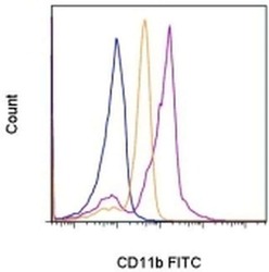
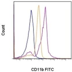
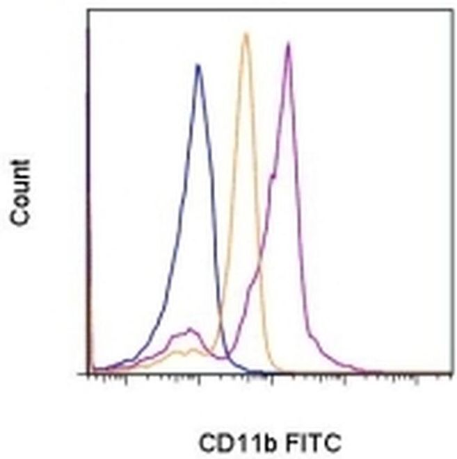
- Staining of 5-minute PMA/Ionomycin-stimulated normal human peripheral blood cells with Mouse IgG1 K Isotype Control FITC (Product # 11-4714-42) (blue histogram) or Anti-Human CD11b (activation epitope) FITC (purple histogram). The orange histogram depicts staining of unstimulated cells with the Anti-Human CD11b (activation epitope) FITC antibody. Cells in the granulocyte gate were used for analysis.
- Conjugate
- Green dye
- Submitted by
- Invitrogen Antibodies (provider)
- Main image
- Experimental details
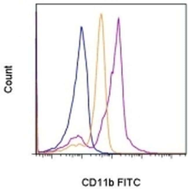
- Staining of 5-minute PMA/Ionomycin-stimulated normal human peripheral blood cells with Mouse IgG1 K Isotype Control FITC (Product # 11-4714-42) (blue histogram) or Anti-Human CD11b (activation epitope) FITC (purple histogram). The orange histogram depicts staining of unstimulated cells with the Anti-Human CD11b (activation epitope) FITC antibody. Cells in the granulocyte gate were used for analysis.
Supportive validation
- Submitted by
- Invitrogen Antibodies (provider)
- Main image
- Experimental details
- NULL
- Conjugate
- Green dye
- Submitted by
- Invitrogen Antibodies (provider)
- Main image
- Experimental details
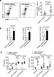
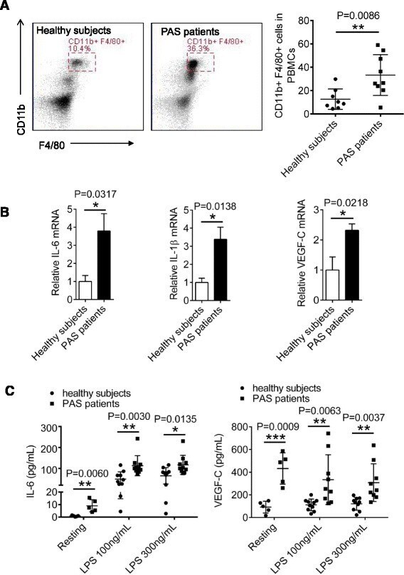
- Fig. 2 Blood monocytes in PAS patients produce inflammatory cytokines and VEGF-C. a PBMCs were isolated from blood of 8 healthy subjects and 9 PAS patients by density-gradient centrifugation with Ficoll (1.077), followed by staining with FITC-conjugated anti-CD11b and APC-conjugated anti-F4/80 for flow cytometer analysis. b Relative mRNA levels IL-6, VEGF-C or IL-1beta in PBMCs from healthy subjects and PAS patients were tested with qRT-PCR. c Freshly isolated monocytes from healthy subjects and PAS patients were either untreated (n = 6) or stimulated with different doses of LPS for 24 h (n = 10), followed by detection of IL-6 and VEGF-C concentrations from the supernatants by ELISA. Data presented are representative of three replicated experiments
- Conjugate
- Green dye
- Submitted by
- Invitrogen Antibodies (provider)
- Main image
- Experimental details
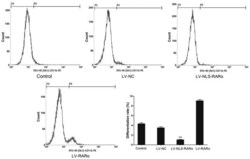
- Figure 3. NLS-RARalpha inhibits the differentiation of NB4 cells. The differentiation rates of each group were detected using flow cytometry. All-trans retinoic acid was used to induce cell differentiation at a concentration of 1 nM. The differentiation rate of cells in the LV-NLS-RARalpha group decreased significantly. **P
- Conjugate
- Green dye

- Submitted by
- Invitrogen Antibodies (provider)
- Main image
- Experimental details
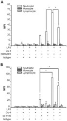
- Figure 4 Effects of Gu-4 on the expression of CD11b and the exposure of CD11b active I-domain. In the presence or absence of Gu-4 (40 nmol/ml), whole blood samples from healthy donors were firstly stimulated with or without LPS (100 ng/ml) for 30 mins, then the samples were co-incubated with saturating amounts of PE conjugated CBRM1/5 or PE labeled anti-CD11b antibody sc-1186 for 10 mins, respectively. After removal of red blood cells, different populations of leukocytes were identified by flow cytometry. A: Expression of CD11b active I-domain. Upon LPS stimulation, CD11b active I-domain on neutrophils was greatly increased, but could be markedly supressed by Gu-4 treatment. B: Expression of CD11b. CD11b expression was significantly elevated in LPS-stimulated neutrophils. Gu-4 showed weak inhibitory effect on such elevation. Data represented the results of triplicate experiments. *: p
- Conjugate
- Green dye

- Submitted by
- Invitrogen Antibodies (provider)
- Main image
- Experimental details
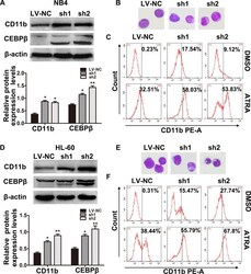
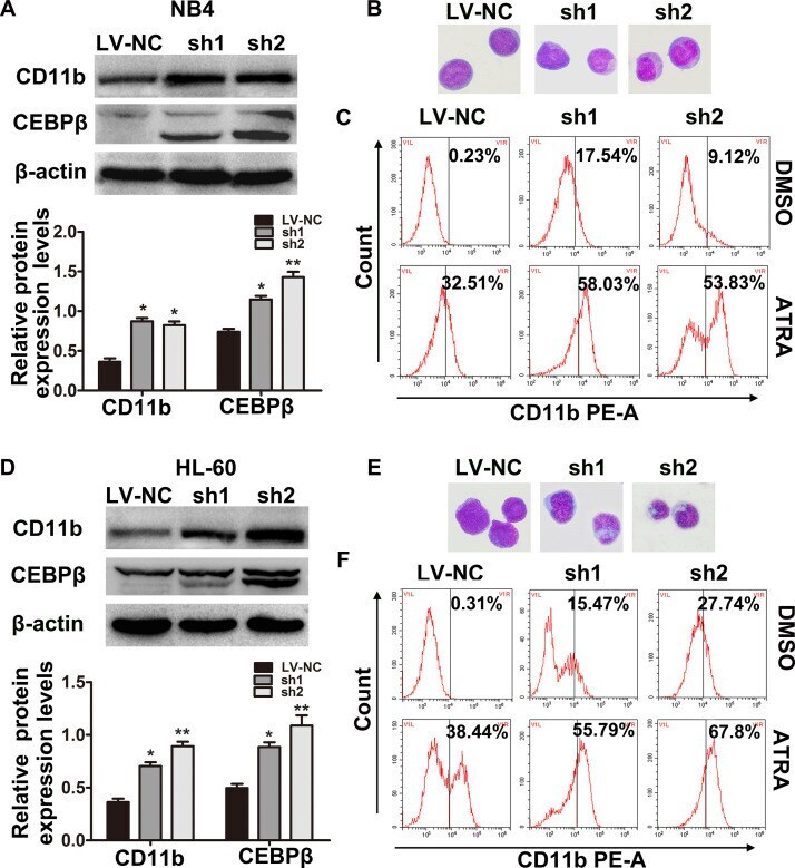
- Figure 4 Downregulation of RREB1 enhances differentiation of NB4 and HL-60 cells. (A) The protein levels of CD11b and CEBPbeta in NB4 (A) and HL-60 (D) cells were examined by Western blot. Representative Wright-Giemsa staining images showed the change in cell morphology in NB4 (B) and HL-60 (E) cells. Original magnification: 100x. The CD11b + cells in NB4 (C) and HL-60 (F) cells were analyzed by flow cytometry. * p < 0.05, ** p < 0.01, compared with LV-NC.
- Conjugate
- Green dye
- Submitted by
- Invitrogen Antibodies (provider)
- Main image

- Experimental details
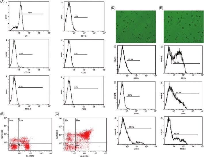
- FIGURE 1 Extraction, identification, and purification of myeloid-derived suppressor cell and cultivation of dendritic cell. (A) Cells were extracted from the bone marrow of BALB/c mice and stained by monoclonal antibodies. Under flow cytometry, the expressive rate of Gr-1 + MDSC, CD11b + MDSC, CD11c + MDSC, CD80 + MDSC, F4/80 + MDSC, and MHC-II + MDSC were 70.4%, 3.5%, 4.8%, 1.2%, 0.3%, 2.1% respectively. (B) The expressive rate of Gr-1 + CD11b + MDSC was 22.6%. (C) After MACS by CD11b magnetic bead, purification of Gr-1 + CD11b + MDSC reached 84.6%. (D) Most non-antigen-loaded dendritic cells grew adherently, with different sizes, star or spindle shape, and stretching tubers, but some of the cells seemed to have adopted a half-adherent state with rough surface. The expressive rates of CD11c, CD86, and MHC-II on DCs were 10.9%, 3.8%, and 27.9%, respectively, by flow cytometry. (E) On the 7th day, DCs were stimulated and activated by tumor antigens. DCs in the half-adherent state increased obviously with radial spikes and bigger shape. The expressive rates of CD11c, CD86, and MHC-II were 74.8%, 50.3%, and 49.8%, respectively, by flow cytometry. IgG FITC, a homotypic control antibody, was used to set the gate strategy. Scale bar = 100 mumol/liter. MDSC, myeloid-derived suppressor cell; MACS, magnetic-activated cell sorting; DC, dendritic cell; CD, cluster of differentiation
- Conjugate
- Green dye
- Submitted by
- Invitrogen Antibodies (provider)
- Main image
- Experimental details
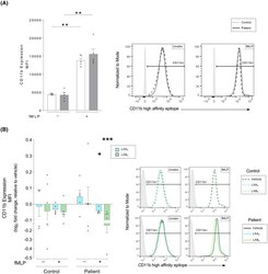
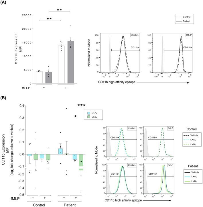
- FIGURE 4 Lipoxin-mediated changes to the high-affinity conformation of the CD11b receptor in human neutrophils from patients with atherosclerosis versus healthy controls. Whole blood from healthy controls ( n = 5) or patients with atherosclerosis ( n = 5) was exposed to inflammatory stimulus as indicated, either in the absence or presence of lipoxin A 4 (LXA 4 : 500 nM) or lipoxin B 4 (LXB 4 : 500 nM). Neutrophil expression of the CD11b high-affinity conformation was measured by flow cytometry. (A) Neutrophil expression of the CD11b high-affinity conformation was measured as the cellular mean fluorescence intensity (MFI). The expression was measured in controls (white bars) and patients (gray bars). The cells were untreated (Unstim.) or stimulated with chemotactic peptide N-formyl-Met-Leu-Phe (fMLP, 0.4 muM). Representative MFI histograms for CD11b expression and respective conditions are shown for controls (dashed line) and patients (solid line), where the gates were determined using a negative population (gray shaded peaks). (B) LXA 4 (blue bars) and LXB 4 (green greens)-induced changes to the neutrophil expression of the CD11b high-affinity conformation was calculated as the log 2 fold change relative to respective vehicle-treated condition. The samples were stimulated as indicated. The bar graphs show levels of cellular CD11b MFI. Representative histograms for the expression of CD11b and respective conditions are shown for vehicle (black line), LXA 4 (blue line), and LXB
- Conjugate
- Green dye
- Submitted by
- Invitrogen Antibodies (provider)
- Main image
- Experimental details
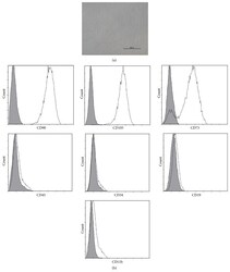
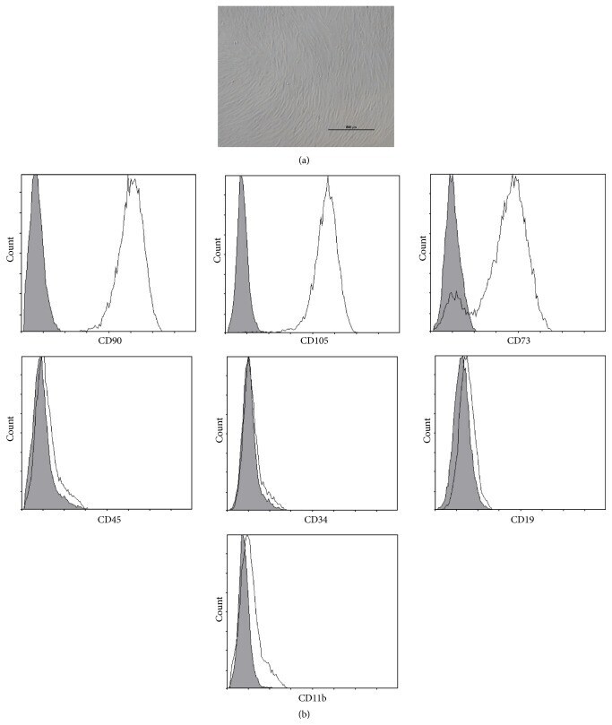
- Figure 1 Characteristics of BM-MSCs. (a) Representative morphology of BM-MSCs. Scale bar = 500 mu m. (b) Representative flow cytometric characterization of cell surface markers expressed on BM-MSCs. Isotypic controls were represented by the gray filled histograms.
- Conjugate
- Green dye
- Submitted by
- Invitrogen Antibodies (provider)
- Main image
- Experimental details
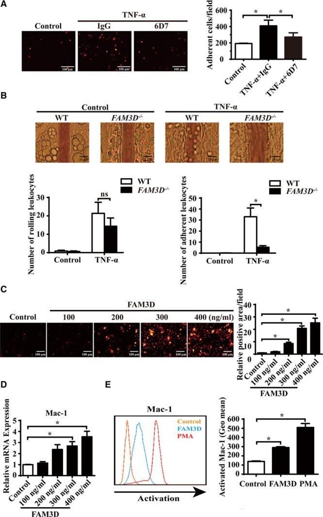
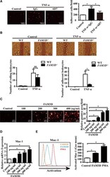
- Figure 5. FAM3D (family with sequence similarity 3, member D) induces neutrophil adhesion and transmigration and activates Mac-1 (macrophage-1 antigen) in neutrophils. A , Representative photographs and quantification of the adhesion of DiI-labeled neutrophils to human umbilical vein endothelial cells (HUVECs) after coculture in the absence or presence of FAM3D neutralization antibody 6D7 (20 mumol/L) or an equal amount of mouse IgG for 2 hours. HUVEC monolayers were stimulated with 4 ng/mL TNF-alpha (tumor necrosis factor alpha) for 24 hours prior to coculture. n=12. One-way analysis of variance (ANOVA) followed by Tukey's test for multiple comparisons, * P
- Conjugate
- Green dye