 Explore
 Validate
 Learn
Learn Immunocytochemistry
Antibody data
- Antibody Data
- Antigen structure
- References [0]
- Comments [0]
- Validations
- Immunocytochemistry [1]
- Immunohistochemistry [1]
- Flow cytometry [1]
Submit
Validation data
Reference
Comment
Report error
- Product number
- MAB16991 - Provider product page

- Provider
- R&D Systems
- Product name
- Human/Equine CD11b/Integrin alpha M Antibody
- Antibody type
- Monoclonal
- Description
- Protein A or G purified from hybridoma culture supernatant. Detects human CD11b/Integrin alpha M in direct ELISAs.
- Reactivity
- Human
- Host
- Mouse
- Conjugate
- Unconjugated
- Antigen sequence
NP_001139280- Isotype
- IgG
- Antibody clone number
- 238446
- Vial size
- 100 ug
- Concentration
- LYOPH
- Storage
- Use a manual defrost freezer and avoid repeated freeze-thaw cycles. 12 months from date of receipt, -20 to -70 °C as supplied. 1 month, 2 to 8 °C under sterile conditions after reconstitution. 6 months, -20 to -70 °C under sterile conditions after reconstitution.
No comments: Submit comment
Supportive validation
- Submitted by
- R&D Systems (provider)
- Main image

- Experimental details
- CD11b/Integrin alpha M in Human PBMCs. CD11b/Integrin alpha M was detected in immersion fixed human peripheral blood mononuclear cells (PBMCs) using Mouse Anti-Human/Equine CD11b/Integrin alpha M Monoclonal Antibody (Catalog # MAB16991) at 10 µg/mL for 3 hours at room temperature. Cells were stained using the Northern-Lights™ 557-conjugated Anti-Mouse IgG Secondary Antibody (red; Catalog # NL007) and counterstained with DAPI (blue). View our protocol for Fluorescent ICC Staining of Non-adherent Cells.

Supportive validation
- Submitted by
- R&D Systems (provider)
- Main image
- Experimental details
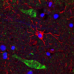
- CD11b/Integrin alpha M in Human Brain. CD11b/Integrin alpha M was detected in immersion fixed paraffin-embedded sections of human brain (cerebral cortex) using Mouse Anti-Human/Equine CD11b/Integrin alpha M Monoclonal Antibody (Catalog # MAB16991). Before incubation with the primary antibody, tissue was subjected to heat-induced epitope retrieval using Antigen Retrieval Reagent-Basic (Catalog # CTS013). Tissue was stained using the NorthernLights™ 557-conjugated Anti-Mouse IgG Secondary Antibody (red; Catalog # NL007) and counterstained with DAPI (blue). Specific staining was localized to cytoplasm of microglia. Tissue was co-stained with using a Sheep Anti-Human/Mouse/Rat Neurogranin Antigen Affinity-Purified Polyclonal Anitbody (Catalog # AF7947) and an Alexa Fluor® 488-conjugated Donkey Anti-Sheep IgG Secondary Antibody (green). View our protocol for Fluorescent IHC Staining of Frozen Tissue Sections.

Supportive validation
- Submitted by
- R&D Systems (provider)
- Main image

- Experimental details
- Detection of CD11b/Integrin alpha M in Equine PBMCs by Flow Cytometry. Equine peripheral blood mononuclear cells (PBMCs) were stained with Mouse Anti-Human/Equine CD11b/Integrin alpha M Monoclonal Antibody (Catalog # MAB16991, filled histogram) or isotype control antibody (Catalog # MAB004, open histogram), followed by Phycoerythrin-conjugated Anti-Mouse IgG Secondary Antibody (Catalog # F0102B).
