Explore
 Validate
 Learn
Learn Western blot
 Immunocytochemistry
Antibody data
- Antibody Data
- Antigen structure
- References [22]
- Comments [0]
- Validations
- Immunocytochemistry [2]
- Immunohistochemistry [1]
- Other assay [13]
Submit
Validation data
Reference
Comment
Report error
- Product number
- PA1-722 - Provider product page

- Provider
- Invitrogen Antibodies
- Product name
- PDE6B Polyclonal Antibody
- Antibody type
- Polyclonal
- Antigen
- Synthetic peptide
- Description
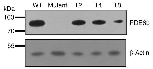
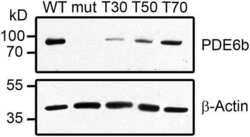
- PA1-722 detects phosphodiesterase 6 beta (PDE6 beta) from mouse retinal extracts. PA1-722 has been successfully used in Western blot procedures. By Western blot, this antibody detects an ~90 kDa protein representing PDE6 beta from mouse retinal extracts. The PA1-722 immunizing peptides correspond to amino acid residues 20-36 from mouse and bovine PDE6 beta protein. These peptides (Cat. # PEP-183) are available for use in neutralization and control experiments.
- Reactivity
- Human, Mouse
- Host
- Rabbit
- Isotype
- IgG
- Vial size
- 100 μg
- Concentration
- 1 mg/mL
- Storage
- -20°C, Avoid Freeze/Thaw Cycles
Submitted references Molecular insights into the maturation of phosphodiesterase 6 by the specialized chaperone complex of HSP90 with AIPL1.
Late-stage rescue of visually guided behavior in the context of a significantly remodeled retinitis pigmentosa mouse model.
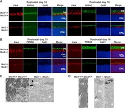
The Musashi proteins MSI1 and MSI2 are required for photoreceptor morphogenesis and vision in mice.
Inhibition of Epigenetic Modifiers LSD1 and HDAC1 Blocks Rod Photoreceptor Death in Mouse Models of Retinitis Pigmentosa.
The ubiquitin-like modifier FAT10 inhibits retinal PDE6 activity and mediates its proteasomal degradation.
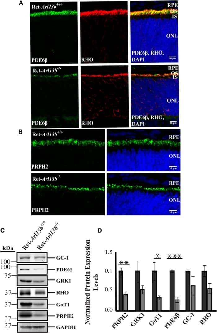
ARL13B, a Joubert Syndrome-Associated Protein, Is Critical for Retinogenesis and Elaboration of Mouse Photoreceptor Outer Segments.
Genetic rescue models refute nonautonomous rod cell death in retinitis pigmentosa.
Halting progressive neurodegeneration in advanced retinitis pigmentosa.
Gene therapy restores vision in rd1 mice after removal of a confounding mutation in Gpr179.
Generation of three-dimensional retinal tissue with functional photoreceptors from human iPSCs.
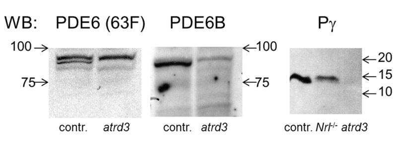
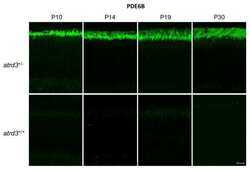
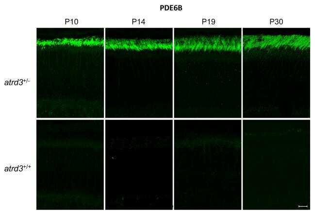
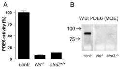
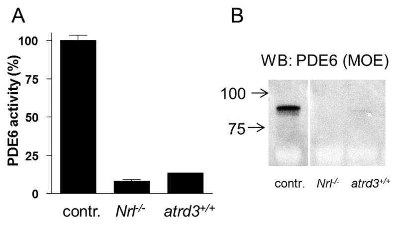
Atypical retinal degeneration 3 in mice is caused by defective PDE6B pre-mRNA splicing.
Ccdc66 null mutation causes retinal degeneration and dysfunction.
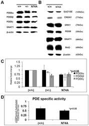
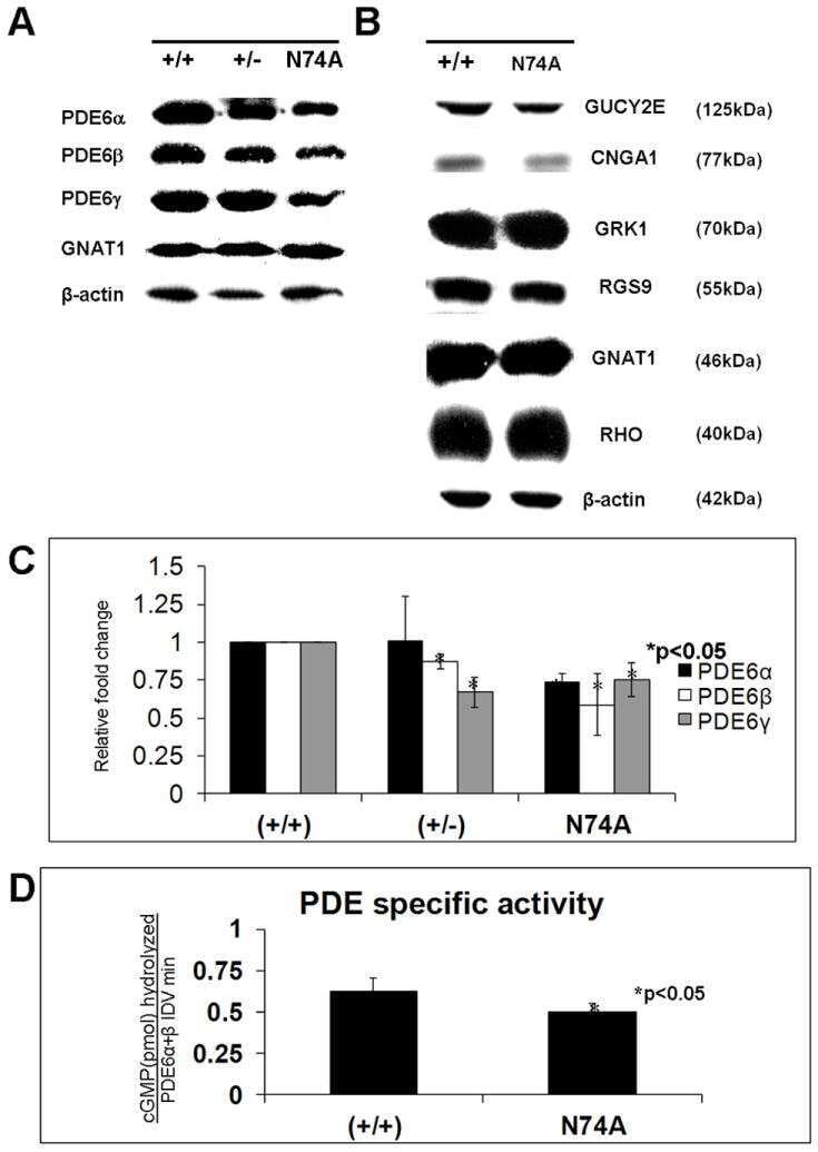
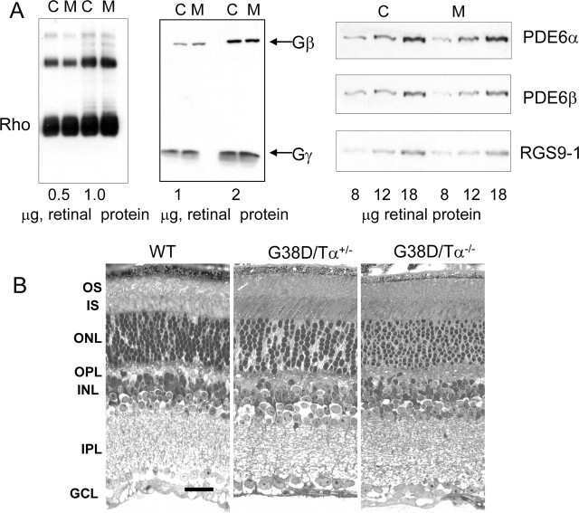
Function of the asparagine 74 residue of the inhibitory γ-subunit of retinal rod cGMP-phophodiesterase (PDE) in vivo.
RAS-converting enzyme 1-mediated endoproteolysis is required for trafficking of rod phosphodiesterase 6 to photoreceptor outer segments.
Long-term retinal function and structure rescue using capsid mutant AAV8 vector in the rd10 mouse, a model of recessive retinitis pigmentosa.
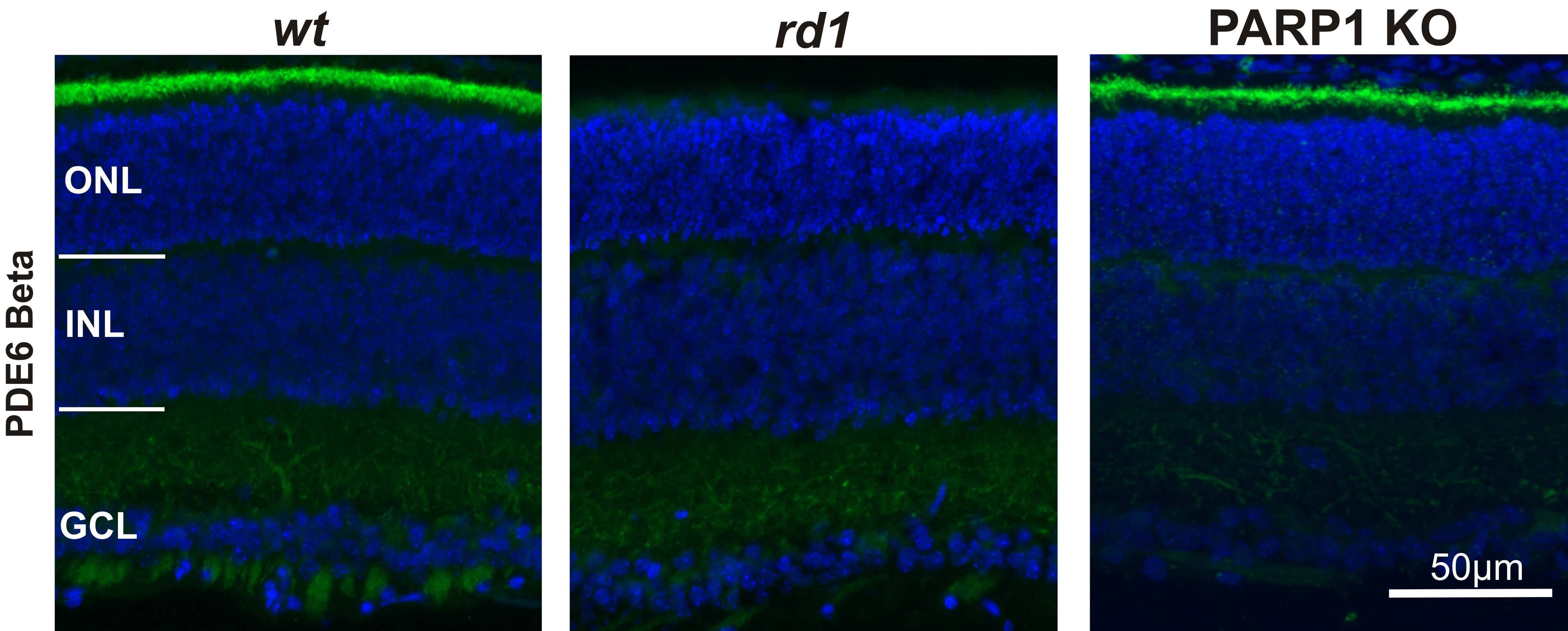
PARP1 gene knock-out increases resistance to retinal degeneration without affecting retinal function.
Functional rescue of degenerating photoreceptors in mice homozygous for a hypomorphic cGMP phosphodiesterase 6 b allele (Pde6bH620Q).
Phototransduction in a transgenic mouse model of Nougaret night blindness.
The inhibitory gamma subunit of the rod cGMP phosphodiesterase binds the catalytic subunits in an extended linear structure.
Transducin activation state controls its light-dependent translocation in rod photoreceptors.
Mammalian sperm phosphodiesterases and their involvement in receptor-mediated cell signaling important for capacitation.
AIPL1, the protein that is defective in Leber congenital amaurosis, is essential for the biosynthesis of retinal rod cGMP phosphodiesterase.
Yadav RP, Boyd K, Artemyev NO
The Journal of biological chemistry 2022 Mar;298(3):101620
The Journal of biological chemistry 2022 Mar;298(3):101620
Late-stage rescue of visually guided behavior in the context of a significantly remodeled retinitis pigmentosa mouse model.
Kajtna J, Tsang SH, Koch SF
Cellular and molecular life sciences : CMLS 2022 Feb 23;79(3):148
Cellular and molecular life sciences : CMLS 2022 Feb 23;79(3):148
The Musashi proteins MSI1 and MSI2 are required for photoreceptor morphogenesis and vision in mice.
Sundar J, Matalkah F, Jeong B, Stoilov P, Ramamurthy V
The Journal of biological chemistry 2021 Jan-Jun;296:100048
The Journal of biological chemistry 2021 Jan-Jun;296:100048
Inhibition of Epigenetic Modifiers LSD1 and HDAC1 Blocks Rod Photoreceptor Death in Mouse Models of Retinitis Pigmentosa.
Popova EY, Imamura Kawasawa Y, Zhang SS, Barnstable CJ
The Journal of neuroscience : the official journal of the Society for Neuroscience 2021 Aug 4;41(31):6775-6792
The Journal of neuroscience : the official journal of the Society for Neuroscience 2021 Aug 4;41(31):6775-6792
The ubiquitin-like modifier FAT10 inhibits retinal PDE6 activity and mediates its proteasomal degradation.
Boehm AN, Bialas J, Catone N, Sacristan-Reviriego A, van der Spuy J, Groettrup M, Aichem A
The Journal of biological chemistry 2020 Oct 16;295(42):14402-14418
The Journal of biological chemistry 2020 Oct 16;295(42):14402-14418
ARL13B, a Joubert Syndrome-Associated Protein, Is Critical for Retinogenesis and Elaboration of Mouse Photoreceptor Outer Segments.
Dilan TL, Moye AR, Salido EM, Saravanan T, Kolandaivelu S, Goldberg AFX, Ramamurthy V
The Journal of neuroscience : the official journal of the Society for Neuroscience 2019 Feb 20;39(8):1347-1364
The Journal of neuroscience : the official journal of the Society for Neuroscience 2019 Feb 20;39(8):1347-1364
Genetic rescue models refute nonautonomous rod cell death in retinitis pigmentosa.
Koch SF, Duong JK, Hsu CW, Tsai YT, Lin CS, Wahl-Schott CA, Tsang SH
Proceedings of the National Academy of Sciences of the United States of America 2017 May 16;114(20):5259-5264
Proceedings of the National Academy of Sciences of the United States of America 2017 May 16;114(20):5259-5264
Halting progressive neurodegeneration in advanced retinitis pigmentosa.
Koch SF, Tsai YT, Duong JK, Wu WH, Hsu CW, Wu WP, Bonet-Ponce L, Lin CS, Tsang SH
The Journal of clinical investigation 2015 Sep;125(9):3704-13
The Journal of clinical investigation 2015 Sep;125(9):3704-13
Gene therapy restores vision in rd1 mice after removal of a confounding mutation in Gpr179.
Nishiguchi KM, Carvalho LS, Rizzi M, Powell K, Holthaus SM, Azam SA, Duran Y, Ribeiro J, Luhmann UF, Bainbridge JW, Smith AJ, Ali RR
Nature communications 2015 Jan 23;6:6006
Nature communications 2015 Jan 23;6:6006
Generation of three-dimensional retinal tissue with functional photoreceptors from human iPSCs.
Zhong X, Gutierrez C, Xue T, Hampton C, Vergara MN, Cao LH, Peters A, Park TS, Zambidis ET, Meyer JS, Gamm DM, Yau KW, Canto-Soler MV
Nature communications 2014 Jun 10;5:4047
Nature communications 2014 Jun 10;5:4047
Atypical retinal degeneration 3 in mice is caused by defective PDE6B pre-mRNA splicing.
Muradov H, Boyd KK, Kerov V, Artemyev NO
Vision research 2012 Mar 15;57:1-8
Vision research 2012 Mar 15;57:1-8
Ccdc66 null mutation causes retinal degeneration and dysfunction.
Gerding WM, Schreiber S, Schulte-Middelmann T, de Castro Marques A, Atorf J, Akkad DA, Dekomien G, Kremers J, Dermietzel R, Gal A, Rülicke T, Ibrahim S, Epplen JT, Petrasch-Parwez E
Human molecular genetics 2011 Sep 15;20(18):3620-31
Human molecular genetics 2011 Sep 15;20(18):3620-31
Function of the asparagine 74 residue of the inhibitory γ-subunit of retinal rod cGMP-phophodiesterase (PDE) in vivo.
Tsang SH, Woodruff ML, Hsu CW, Naumann MC, Cilluffo M, Tosi J, Lin CS
Cellular signalling 2011 Oct;23(10):1584-9
Cellular signalling 2011 Oct;23(10):1584-9
RAS-converting enzyme 1-mediated endoproteolysis is required for trafficking of rod phosphodiesterase 6 to photoreceptor outer segments.
Christiansen JR, Kolandaivelu S, Bergo MO, Ramamurthy V
Proceedings of the National Academy of Sciences of the United States of America 2011 May 24;108(21):8862-6
Proceedings of the National Academy of Sciences of the United States of America 2011 May 24;108(21):8862-6
Long-term retinal function and structure rescue using capsid mutant AAV8 vector in the rd10 mouse, a model of recessive retinitis pigmentosa.
Pang JJ, Dai X, Boye SE, Barone I, Boye SL, Mao S, Everhart D, Dinculescu A, Liu L, Umino Y, Lei B, Chang B, Barlow R, Strettoi E, Hauswirth WW
Molecular therapy : the journal of the American Society of Gene Therapy 2011 Feb;19(2):234-42
Molecular therapy : the journal of the American Society of Gene Therapy 2011 Feb;19(2):234-42
PARP1 gene knock-out increases resistance to retinal degeneration without affecting retinal function.
Sahaboglu A, Tanimoto N, Kaur J, Sancho-Pelluz J, Huber G, Fahl E, Arango-Gonzalez B, Zrenner E, Ekström P, Löwenheim H, Seeliger M, Paquet-Durand F
PloS one 2010 Nov 23;5(11):e15495
PloS one 2010 Nov 23;5(11):e15495
Functional rescue of degenerating photoreceptors in mice homozygous for a hypomorphic cGMP phosphodiesterase 6 b allele (Pde6bH620Q).
Davis RJ, Tosi J, Janisch KM, Kasanuki JM, Wang NK, Kong J, Tsui I, Cilluffo M, Woodruff ML, Fain GL, Lin CS, Tsang SH
Investigative ophthalmology & visual science 2008 Nov;49(11):5067-76
Investigative ophthalmology & visual science 2008 Nov;49(11):5067-76
Phototransduction in a transgenic mouse model of Nougaret night blindness.
Moussaif M, Rubin WW, Kerov V, Reh R, Chen D, Lem J, Chen CK, Hurley JB, Burns ME, Artemyev NO
The Journal of neuroscience : the official journal of the Society for Neuroscience 2006 Jun 21;26(25):6863-72
The Journal of neuroscience : the official journal of the Society for Neuroscience 2006 Jun 21;26(25):6863-72
The inhibitory gamma subunit of the rod cGMP phosphodiesterase binds the catalytic subunits in an extended linear structure.
Guo LW, Muradov H, Hajipour AR, Sievert MK, Artemyev NO, Ruoho AE
The Journal of biological chemistry 2006 Jun 2;281(22):15412-22
The Journal of biological chemistry 2006 Jun 2;281(22):15412-22
Transducin activation state controls its light-dependent translocation in rod photoreceptors.
Kerov V, Chen D, Moussaif M, Chen YJ, Chen CK, Artemyev NO
The Journal of biological chemistry 2005 Dec 9;280(49):41069-76
The Journal of biological chemistry 2005 Dec 9;280(49):41069-76
Mammalian sperm phosphodiesterases and their involvement in receptor-mediated cell signaling important for capacitation.
Baxendale RW, Fraser LR
Molecular reproduction and development 2005 Aug;71(4):495-508
Molecular reproduction and development 2005 Aug;71(4):495-508
AIPL1, the protein that is defective in Leber congenital amaurosis, is essential for the biosynthesis of retinal rod cGMP phosphodiesterase.
Liu X, Bulgakov OV, Wen XH, Woodruff ML, Pawlyk B, Yang J, Fain GL, Sandberg MA, Makino CL, Li T
Proceedings of the National Academy of Sciences of the United States of America 2004 Sep 21;101(38):13903-8
Proceedings of the National Academy of Sciences of the United States of America 2004 Sep 21;101(38):13903-8
No comments: Submit comment
Supportive validation
- Submitted by
- Invitrogen Antibodies (provider)
- Main image

- Experimental details
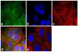
- Immunofluorescence analysis of PDE6 beta was performed using 70% confluent log phase MCF-7 cells. The cells were fixed with 4% paraformaldehyde for 10 minutes, permeabilized with 0.1% Triton™ X-100 for 10 minutes, and blocked with 1% BSA for 1 hour at room temperature. The cells were labeled with PDE6B Rabbit Polyclonal Antibody (Product # PA1-722) at 1:250 dilution in 0.1% BSA and incubated for 3 hours at room temperature and then labeled with Goat anti-Rabbit IgG (H+L) Superclonal™ Secondary Antibody, Alexa Fluor® 488 conjugate (Product # A27034) at a dilution of 1:2000 for 45 minutes at room temperature (Panel a: green). Nuclei (Panel b: blue) were stained with SlowFade® Gold Antifade Mountant with DAPI (Product # S36938). F-actin (Panel c: red) was stained with Rhodamine Phalloidin (Product # R415, 1:300). Panel d represents the merged image showing cytoplasmic and nuclear localization. Panel e shows the no primary antibody control. The images were captured at 60X magnification.

- Submitted by
- Invitrogen Antibodies (provider)
- Main image
- Experimental details
- Immunofluorescence analysis of PDE6 beta was performed using 70% confluent log phase MCF-7 cells. The cells were fixed with 4% paraformaldehyde for 10 minutes, permeabilized with 0.1% Triton™ X-100 for 10 minutes, and blocked with 1% BSA for 1 hour at room temperature. The cells were labeled with PDE6B Rabbit Polyclonal Antibody (Product # PA1-722) at 1:250 dilution in 0.1% BSA and incubated for 3 hours at room temperature and then labeled with Goat anti-Rabbit IgG (Heavy Chain) Superclonal™ Secondary Antibody, Alexa Fluor® 488 conjugate (Product # A27034) at a dilution of 1:2000 for 45 minutes at room temperature (Panel a: green). Nuclei (Panel b: blue) were stained with SlowFade® Gold Antifade Mountant with DAPI (Product # S36938). F-actin (Panel c: red) was stained with Rhodamine Phalloidin (Product # R415, 1:300). Panel d represents the merged image showing cytoplasmic and nuclear localization. Panel e shows the no primary antibody control. The images were captured at 60X magnification.

Supportive validation
- Submitted by
- Invitrogen Antibodies (provider)
- Main image
- Experimental details
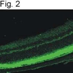
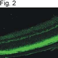
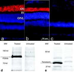
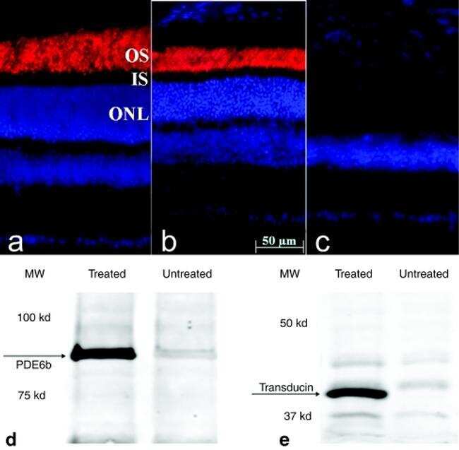
- Immunofluorscences of mouse retina outer segments after staining with PA1-722.
Supportive validation
- Submitted by
- Invitrogen Antibodies (provider)
- Main image
- Experimental details
- NULL
- Submitted by
- Invitrogen Antibodies (provider)
- Main image
- Experimental details
- NULL
- Submitted by
- Invitrogen Antibodies (provider)
- Main image
- Experimental details
- NULL
- Submitted by
- Invitrogen Antibodies (provider)
- Main image

- Experimental details
- NULL
- Submitted by
- Invitrogen Antibodies (provider)
- Main image
- Experimental details
- NULL
- Submitted by
- Invitrogen Antibodies (provider)
- Main image
- Experimental details
- NULL
- Submitted by
- Invitrogen Antibodies (provider)
- Main image
- Experimental details
- NULL

- Submitted by
- Invitrogen Antibodies (provider)
- Main image

- Experimental details
- NULL
- Submitted by
- Invitrogen Antibodies (provider)
- Main image
- Experimental details
- NULL
- Submitted by
- Invitrogen Antibodies (provider)
- Main image
- Experimental details
- NULL

- Submitted by
- Invitrogen Antibodies (provider)
- Main image

- Experimental details
- NULL
- Submitted by
- Invitrogen Antibodies (provider)
- Main image
- Experimental details
- NULL
- Submitted by
- Invitrogen Antibodies (provider)
- Main image
- Experimental details
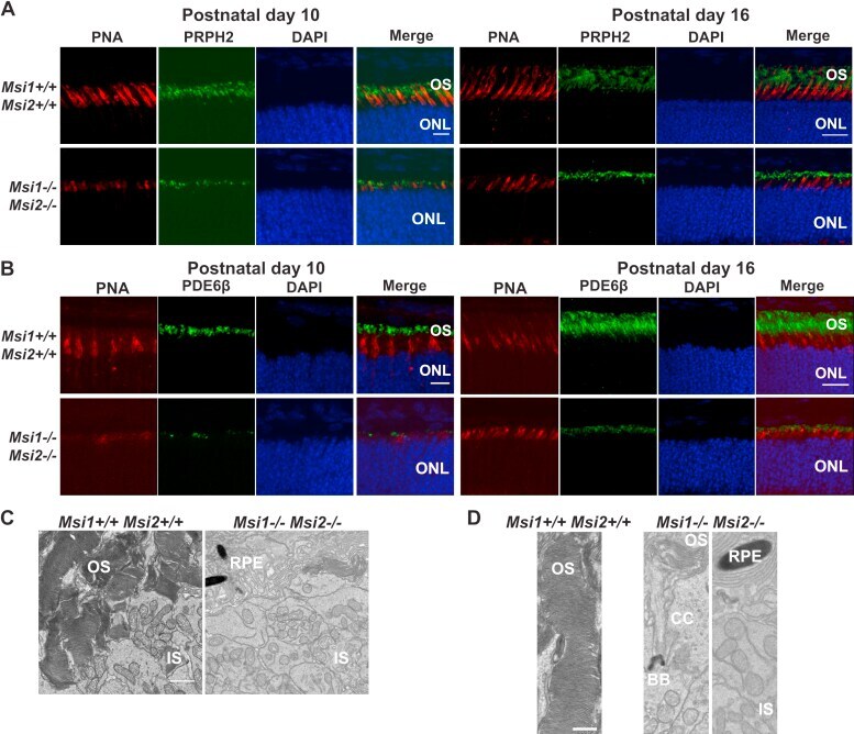
- Figure 6 Abnormal development of OS in the absence of MSI1 and MSI2. A , immunofluorescence microscopy images of retinal cross sections from the ret-Msi1-/-: Msi2-/- mice at P10 ( left ) and P16 ( right ) stained with anti-peripherin-2 antibody (PRPH2: OS marker- green ) and peanut agglutinin (PNA: cone OS marker- red ) along with a DAPI nuclear counterstain ( blue ). Scale bar = 20 mum. B , immunofluorescence microscopy images of retinal cross sections from the ret-Msi1-/-: Msi2-/- mice at P10 ( left ) and P16 ( right ) stained with anti-phosphodiesterase-6beta antibody (PDE6beta: rod OS marker- green ) and peanut agglutinin (PNA: cone OS marker- red ) along with a DAPI counterstain ( blue ). Scale bar = 20 mum. C , low-magnification transmission electron microscopy images of ultrathin retinal sections from ret-Msi1-/-: Msi2-/- mice at P10 visualizing the boundary between the OS and IS showing the lack of typical outer segments in the absence of the Musashi proteins. Scale bar = 2 mum. D , high-magnification transmission electron microscopy images of ultrathin retinal sections from ret-Msi1-/-: Msi2-/- mice at P10 visualizing the boundary between the OS and IS showing that the OS either does not form ( far right ) or is dysmorphic ( middle ) in the absence of the Musashi proteins. Scale bar = 1 mum. BB, basal body; CC, connecting cilium; DAPI, 4'',6-diamidino-2-phenylindole; IS, inner segment; ONL, outer nuclear layer; OS, outer segment; RPE, retinal pigment epithelium; P, p