 Explore
 Validate
 Learn
Learn Immunocytochemistry
Antibody data
- Antibody Data
- Antigen structure
- References [0]
- Comments [0]
- Validations
- Immunocytochemistry [3]
Submit
Validation data
Reference
Comment
Report error
- Product number
- 710676 - Provider product page

- Provider
- Invitrogen Antibodies
- Product name
- Phospho-IKK alpha/beta (Ser176, Ser180) Recombinant Polyclonal Antibody (7HCLC)
- Antibody type
- Polyclonal
- Antigen
- Synthetic peptide
- Description
- This antibody is predicted to react with Dog, Rabbit, Pig, Mouse, Rat, Bovine and Goat.
- Antibody clone number
- 7HCLC
- Concentration
- 0.5 mg/mL
No comments: Submit comment
Supportive validation
- Submitted by
- Invitrogen Antibodies (provider)
- Main image
- Experimental details
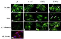
- Time course showing induction of the TNF-a signaling cascade upon treatment: Cellular localization of proteins in the NF-KB signaling pathway was detected upon treatment of HeLa cells with TNF alpha (50 ng/mL) for 5, 10 and 25 min. Fixed and permeabilized cells were stained with Anti-IKK total Recombinant Rabbit Polyclonal Antibody (Product # 710719, 1 µg/mL) or Anti-IKK alpha/beta (pS176/pS180) Recombinant Rabbit Polyclonal Antibody (Product # 710676, 1 µg/mL) or Anti-NF-kB Recombinant Rabbit Polyclonal Antibody (Product # 710048, 1 µg/mL) and labeled with Goat anti-Rabbit IgG (H+L) Superclonal Secondary Antibody, Alexa Fluor® 488 conjugate (Product # A27034, 0.4 µg/mL, 1:2500). Images showing cytoplasmic staining of IKK total and NF-kB (panel a & e; green) in untreated cells. No significant basal levels of phospho-IKK alpha/beta (panel i; green) were detected. Treatment with TNF-a led to a significant increase in the levels of IKK (panel b-d; green) and IKK alpha/beta (pS176/pS180) (panel j-l; green) in the cytosol, specifically at the perinucleus, and a corresponding translocation of NF-KB to the nucleus (panel f-h; green). No background staining was observed in control cells with no primary antibody (panel m).The nuclei (blue) were stained using SlowFade® Gold Antifade Mountant with DAPI (Product # S36938, 1:50). Cytoskeletal F-actin staining (red) was done using Alexa Fluor® 594 Phalloidin (Product # A12381, 1:200).

- Submitted by
- Invitrogen Antibodies (provider)
- Main image
- Experimental details
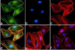
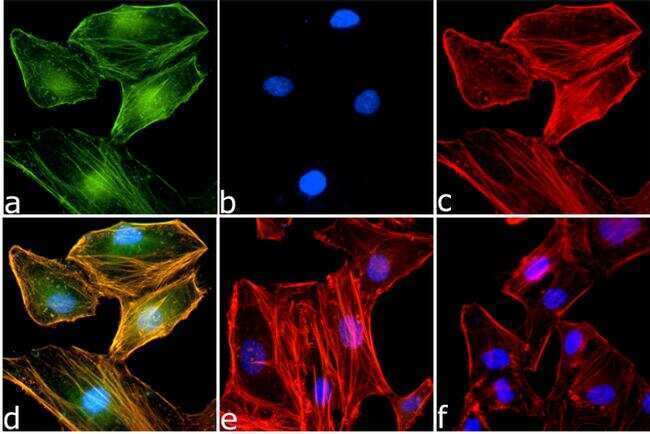
- Immunofluorescence was performed on HeLa cells treated with TNF alpha at 50 ng/mL/25 min. These cells were fixed and permeabilized for detection of IKK alpha/beta (pSpS 176/180) using Anti-IKK alpha/beta (pSpS 176/180) Recombinant Rabbit Polyclonal Antibody (Product # 710676, 1 µg/mL) and labeled with Goat anti-Rabbit IgG (H+L) Superclonal Secondary Antibody, Alexa Fluor® 488 conjugate (Product # A27034, 0.4 µg/mL, 1:2500). Panel a) shows representative cells that were stained for detection and localization protein (green), Panel b) is stained for nuclei (blue) using SlowFade® Gold Antifade Mountant with DAPI (Product # S36938, 1:50). Panel c) represents cytoskeletal F-actin staining using Alexa Fluor® 594 Phalloidin (Product # A12381, 1:200). Panel d) is a composite image of Panels a, b and c clearly demonstrating cytoplasmic and peri-nuclear localization of IKK alpha/beta (pSpS 176/180). Panel e represents control cells with no primary Antibody to assess background. Panel f) shows untreated cells.
- Submitted by
- Invitrogen Antibodies (provider)
- Main image
- Experimental details
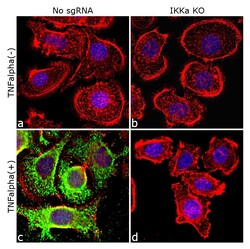
- Immunofluorescence analysis of Phospho-IKK alpha/beta (Ser176, Ser180) was done on 70% confluent log phase ME-180 cells. The cells were either mock treated or treated with TNF-alpha (50 ng/mL for 20 min), fixed with 4% paraformaldehyde for 15 minutes, permeabilized with 0.1% Triton™ X-100 for 10 minutes, and blocked with 1% BSA for 1 hour at room temperature. The cells were subsequently labeled with Phospho-IKK alpha/beta (Ser176, Ser180 - Green) Recombinant Rabbit Polyclonal Antibody (Product # 710676) at 2µg/mL in 0.1% BSA and incubated for 3 hours at room temperature and then labeled with Goat anti-Rabbit IgG (H+L) Superclonal™ Secondary Antibody, Alexa Fluor® 488 conjugate (Product # A27034) at a dilution of 1:2000 for 45 minutes at room temperature. Nuclei (Blue) were stained with SlowFade® Gold Antifade Mountant DAPI (Product # S36938). F-actin (Red) was stained with Rhodamine Phalloidin (Product # R415, 1:300). TNFalpha induced phosphorylation of IKK alpha/beta, was observed in control cell line (panels a, c) and not in the IKK alpha knockout (KO) cell line (panels b, d). The images were captured at 40X magnification.
