Explore
 Validate
 Learn
Learn Western blot
 Immunocytochemistry
Antibody data
- Antibody Data
- Antigen structure
- References [5]
- Comments [0]
- Validations
- Immunocytochemistry [4]
- Immunoprecipitation [1]
- Immunohistochemistry [1]
- Flow cytometry [1]
- Other assay [3]
Submit
Validation data
Reference
Comment
Report error
- Product number
- 700360 - Provider product page

- Provider
- Invitrogen Antibodies
- Product name
- MDA5 Recombinant Rabbit Monoclonal Antibody (33H12L34)
- Antibody type
- Monoclonal
- Antigen
- Recombinant full-length protein
- Description
- This antibody is predicted to react with Rhesus monkey, mouse, rat, porcine and bovine based on sequence homology. Intact IgG appears on a non-reducing gel as ~150 kDa band and upon reduction generating a ~25 kDa light chain band and a ~50 kDa heavy chain. Recombinant rabbit monoclonal antibodies are produced using in vitro expression systems. The expression systems are developed by cloning in the specific antibody DNA sequences from immunoreactive rabbits. Then, individual clones are screened to select the best candidates for production. The advantages of using recombinant rabbit monoclonal antibodies include: better specificity and sensitivity, lot-to-lot consistency, animal origin-free formulations, and broader immunoreactivity to diverse targets due to larger rabbit immune repertoire.
- Reactivity
- Human, Mouse
- Host
- Rabbit
- Isotype
- IgG
- Antibody clone number
- 33H12L34
- Vial size
- 100 μg
- Concentration
- 0.5 mg/mL
- Storage
- Store at 4°C short term. For long term storage, store at -20°C, avoiding freeze/thaw cycles.
Submitted references Dietary Supplementation with Biobran/MGN-3 Increases Innate Resistance and Reduces the Incidence of Influenza-like Illnesses in Elderly Subjects: A Randomized, Double-Blind, Placebo-Controlled Pilot Clinical Trial.
Activation of NF-κB signaling via cytosolic mitochondrial RNA sensing in kerotocytes with mitochondrial DNA common deletion.
Double-stranded RNA innate immune response activation from long-term adeno-associated virus vector transduction.
Rabies Virus Infection Induces the Formation of Stress Granules Closely Connected to the Viral Factories.
HepG2 cells mount an effective antiviral interferon-lambda based innate immune response to hepatitis C virus infection.
Elsaid AF, Agrawal S, Agrawal A, Ghoneum M
Nutrients 2021 Nov 19;13(11)
Nutrients 2021 Nov 19;13(11)
Activation of NF-κB signaling via cytosolic mitochondrial RNA sensing in kerotocytes with mitochondrial DNA common deletion.
Zhou X, Backman LJ, Danielson P
Scientific reports 2021 Apr 1;11(1):7360
Scientific reports 2021 Apr 1;11(1):7360
Double-stranded RNA innate immune response activation from long-term adeno-associated virus vector transduction.
Shao W, Earley LF, Chai Z, Chen X, Sun J, He T, Deng M, Hirsch ML, Ting J, Samulski RJ, Li C
JCI insight 2018 Jun 21;3(12)
JCI insight 2018 Jun 21;3(12)
Rabies Virus Infection Induces the Formation of Stress Granules Closely Connected to the Viral Factories.
Nikolic J, Civas A, Lama Z, Lagaudrière-Gesbert C, Blondel D
PLoS pathogens 2016 Oct;12(10):e1005942
PLoS pathogens 2016 Oct;12(10):e1005942
HepG2 cells mount an effective antiviral interferon-lambda based innate immune response to hepatitis C virus infection.
Israelow B, Narbus CM, Sourisseau M, Evans MJ
Hepatology (Baltimore, Md.) 2014 Oct;60(4):1170-9
Hepatology (Baltimore, Md.) 2014 Oct;60(4):1170-9
No comments: Submit comment
Supportive validation
- Submitted by
- Invitrogen Antibodies (provider)
- Main image

- Experimental details
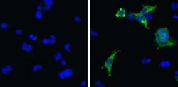
- Immunofluorescent analysis of MDA5 in MDA5-transfected 293 cells (right) and untransfected cells (left) using a MDA5 recombinant rabbit monoclonal antibody (Product # 700360) at a dilution of 5 µg/mL followed by detection using an Alexa Fluor 488-conjugated goat anti-rabbit secondary antibody at a dilution of 1:1000 (green) and nuclei stained using Hoescht (blue).

- Submitted by
- Invitrogen Antibodies (provider)
- Main image
- Experimental details
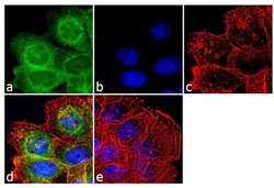
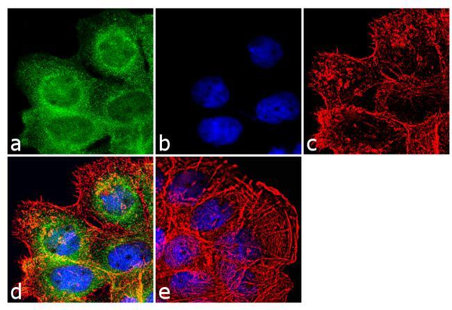
- Immunofluorescence analysis of MDA5 was done on 70% confluent log phase A431 cells. The cells were fixed with 4% paraformaldehyde for 10 minutes, permeabilized with 0.1% Triton™ X-100 for 10 minutes, and blocked with 1% BSA for 1 hour at room temperature. The cells were labeled with MDA5 (33H12L34), Recombinant Rabbit Monoclonal Antibody (Product # 700360) at 2 µg/mL in 0.1% BSA and incubated for 3 hours at room temperature and then labeled with Goat anti-Rabbit IgG (H+L) Superclonal™ Secondary Antibody, Alexa Fluor® 488 conjugate (Product # A27034) at a dilution of 1:2000 for 45 minutes at room temperature (Panel a: green). Nuclei (Panel b: blue) were stained with SlowFade® Gold Antifade Mountant with DAPI (Product # S36938). F-actin (Panel c: red) was stained with Alexa Fluor® 555 Rhodamine Phalloidin (Product # R415, 1:300). Panel d is a merged image showing cytoplasmic and nuclear localization. Panel e is a no primary antibody control. The images were captured at 60X magnification.
- Submitted by
- Invitrogen Antibodies (provider)
- Main image

- Experimental details
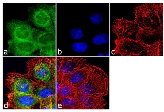
- Immunofluorescence analysis of MDA5 was done on 70% confluent log phase A431 cells. The cells were fixed with 4% paraformaldehyde for 10 minutes, permeabilized with 0.1% Triton™ X-100 for 10 minutes, and blocked with 1% BSA for 1 hour at room temperature. The cells were labeled with MDA5 (33H12L34), Recombinant Rabbit Monoclonal Antibody (Product # 700360) at 2 µg/mL in 0.1% BSA and incubated for 3 hours at room temperature and then labeled with Goat anti-Rabbit IgG (Heavy Chain) Superclonal™ Secondary Antibody, Alexa Fluor® 488 conjugate (Product # A27034) at a dilution of 1:2000 for 45 minutes at room temperature (Panel a: green). Nuclei (Panel b: blue) were stained with SlowFade® Gold Antifade Mountant with DAPI (Product # S36938). F-actin (Panel c: red) was stained with Alexa Fluor® 555 Rhodamine Phalloidin (Product # R415, 1:300). Panel d is a merged image showing cytoplasmic and nuclear localization. Panel e is a no primary antibody control. The images were captured at 60X magnification.
Supportive validation
- Submitted by
- Invitrogen Antibodies (provider)
- Main image
- Experimental details
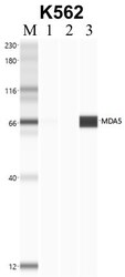
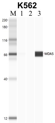
- Immunoprecipitation of MDA5 was performed in K562 cells. Antigen-antibody complexes were formed by incubating approximately 500 µg whole cell lysate with 5 to 10 µL of recombinant monoclonal MDA5 antibody (Product # 700360) rotating 60 min at RT. The immune complexes were captured on 625 µg of anti- rabbit coated Dynabeads (Product # 11204D) and washed extensively. They were then eluted and analyzed using the Simple Western system using the same antibody as used in immunoprecipitation at a dilution of 1:25, followed by a 1:100 dilution of secondary antibody. Lane 1 is the input, lane 2 no antibody IP and lane 3 is the target specific IP. Data courtesy of the Yeo lab as part of the ENCODE project.

Supportive validation
- Submitted by
- Invitrogen Antibodies (provider)
- Main image

- Experimental details
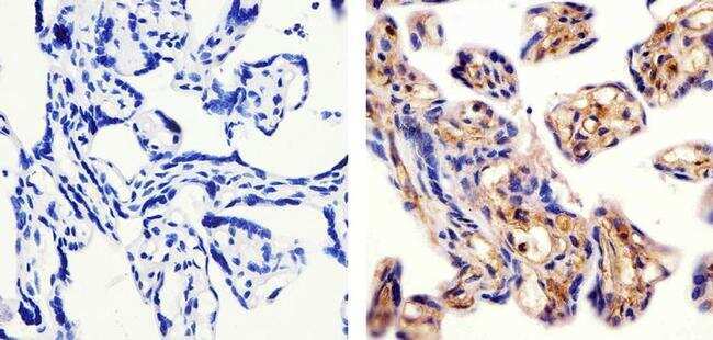
- Immunohistochemistry analysis of MDA5 showing staining in the cytoplasm of paraffin-embedded human placenta tissue (right) compared to a negative control without primary antibody (left). To expose target proteins, antigen retrieval was performed using 10mM sodium citrate (pH 6.0), microwaved for 8-15 min. Following antigen retrieval, tissues were blocked in 3% H2O2-methanol for 15 min at room temperature, washed with ddH2O and PBS, and then probed with a Anti- MDA5 Monoclonal Antibody (Product # 700360) diluted in 3% BSA-PBS at a dilution of 1:20 overnight at 4°C in a humidified chamber. Tissues were washed extensively in PBST and detection was performed using an HRP-conjugated secondary antibody followed by colorimetric detection using a DAB kit. Tissues were counterstained with hematoxylin and dehydrated with ethanol and xylene to prep for mounting.
Supportive validation
- Submitted by
- Invitrogen Antibodies (provider)
- Main image
- Experimental details
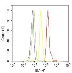
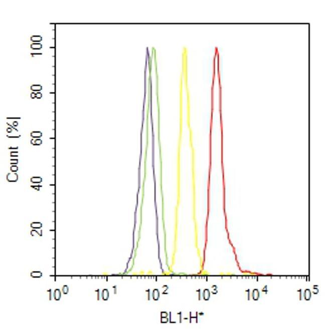
- Flow cytometry analysis of MDA5 was done on MCF7 cells. Cells were fixed with 70% ethanol for 10 minutes, permeabilized with 0.25% Triton™ X-100 for 20 minutes, and blocked with 5% BSA for 30 minutes at room temperature. Cells were labeled with MDA5 Rabbit Monoclonal Antibody (700360, red histogram) or with rabbit isotype control (yellow histogram) at 3-5 ug/million cells in 2.5% BSA. After incubation at room temperature for 2 hours, the cells were labeled with Alexa Fluor® 488 Goat Anti-Rabbit Secondary Antibody (A11008) at a dilution of 1:400 for 30 minutes at room temperature. The representative 10,000 cells were acquired and analyzed for each sample using an Attune® Acoustic Focusing Cytometer. The purple histogram represents unstained control cells and the green histogram represents no-primary-antibody control.
Supportive validation
- Submitted by
- Invitrogen Antibodies (provider)
- Main image

- Experimental details
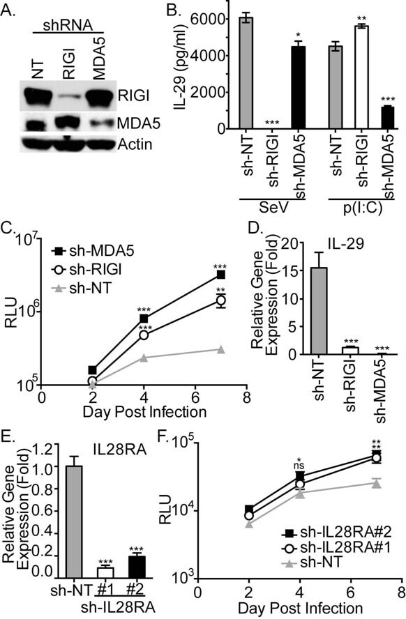
- RIG-I like receptor and IFN-lambda receptor silencing promotes HCV multicycle growth in HepG2-HFL cells. (A) Immunoblots of endogenous RIG-I, MDA5, or ß-actin were performed on lysates from HepG2-HFL cells that were transduced with lentiviral vectors expressing shRNA targeting RIG-I, MDA5, or a no targeting (NT) shRNA used as a negative control. (B) IL-29 production was assayed by ELISA 24 hpi with SeV and 24 hours transfectionwith poly(I:C). (C) HCV-GLuc multicycle growth was monitored by sampling luciferase production at 2, 4, and 7 dpi. (D) IL-29 mRNA induction was assessed in HepG2-HFL cells 30 hpi with non-reporter HCV. (E) Quantification of IFN-lambda receptor (IL28RA) mRNA levels in HepG2-HFL cells transduced with two different lentiviral vectors expressing shRNA targeting IL28RA (#1 and #2), or a control shRNA (NT). (F) HCV-GLuc multicycle growth was monitored by sampling luciferase production at 2, 4, and 7 dpi. Asterisks represent statistically significant differences between NT-shRNA expressing cells and cells expressing shRNA targeting RIG-I, MDA5, or IL-28RA at the indicated time points.
- Submitted by
- Invitrogen Antibodies (provider)
- Main image

- Experimental details
- Immunoprecipitation of MDA5 was performed in K562 cells. Antigen-antibody complexes were formed by incubating approximately 500 µg whole cell lysate with 5 to 10 µL of recombinant monoclonal MDA5 antibody (Product # 700360) rotating 60 min at RT. The immune complexes were captured on 625 µg of anti- rabbit coated Dynabeads (Product # 11204D) and washed extensively. They were then eluted and analyzed using the Simple Western system using the same antibody as used in immunoprecipitation at a dilution of 1:25, followed by a 1:100 dilution of secondary antibody. Lane 1 is the input, lane 2 no antibody IP and lane 3 is the target specific IP. Data courtesy of the Yeo lab as part of the ENCODE project.
- Submitted by
- Invitrogen Antibodies (provider)
- Main image
- Experimental details
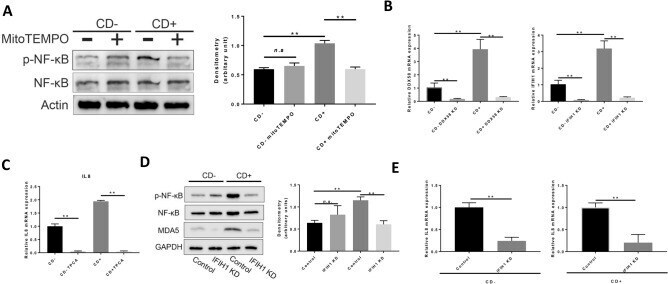
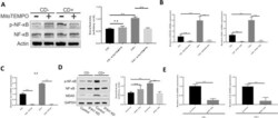
- Figure 2 MDA5 signaling induces IL-8 upregulation via NF-kappaB phosphorylation in CD+ keratocytes. ( A ) Western blot of NF-kappaB phosphorylation in CD- and CD+ keratocytes with/without mitoTEMPO ( n = 4). NF-kappaB and p-NF-kappaB were blotted and cropped from the same gel. p-NF-kappaB was blotted first and the membrane was stripped and washed for NF-kappaB blotting. Actin was blotted separately from a different gel using the corresponding samples. Exposures were adjusted automatically by the Odyssey Fc imaging system. ( B ) DDX58 and IFIH1 mRNA expression in CD+/- keratocytes 48 h after respective siRNA transfection ( n = 3). ( C ) IL-8 mRNA expression in keratocytes 24 h after 20 nM TPCA treatment ( n = 3). ( D ) NF-kappaB phosphorylation, analyzed by western blot, after IFIH1 knock-down in CD+/- keratocytes ( n = 3). NF-kappaB and p-NF-kappaB were blotted and cropped from the same gel. p-NF-kappaB was blotted first and the membrane was stripped and washed for NF-kappaB blotting. GAPDH and MDA5 were blotted separately from a different gel using the corresponding samples. Exposures were adjusted automatically by the Odyssey Fc imaging system. ( E ) IL-8 mRNA expression in CD+/- keratocytes after IFIH1 knockdown by siRNA transfection ( n = 3). Values are means +- SD. n.s. (not significant); *P < 0.05, **P < 0.01.