 Explore
 Validate
 Learn
Learn Flow cytometry
Antibody data
- Antibody Data
- Antigen structure
- References [2]
- Comments [0]
- Validations
- Flow cytometry [1]
Submit
Validation data
Reference
Comment
Report error
- Product number
- IC1935A - Provider product page

- Provider
- Novus Biologicals
- Product name
- Mouse Monoclonal HIF-1 alpha Antibody
- Antibody type
- Monoclonal
- Description
- Protein A or G purified from hybridoma culture supernatant. Recognizes human and mouse HIF-1 alpha in direct ELISAs.
- Reactivity
- Human, Mouse
- Host
- Mouse
- Conjugate
- Red dye
- Isotype
- IgG
- Vial size
- 100 Tests
- Storage
- Protect from light. Do not freeze. 12 months from date of receipt, 2 to 8 degreesC as supplied.
Submitted references Endogenous oxidized phospholipids reprogram cellular metabolism and boost hyperinflammation.
E3 Ligase VHL Promotes Group 2 Innate Lymphoid Cell Maturation and Function via Glycolysis Inhibition and Induction of Interleukin-33 Receptor.
Di Gioia M, Spreafico R, Springstead JR, Mendelson MM, Joehanes R, Levy D, Zanoni I
Nature immunology 2020 Jan;21(1):42-53
Nature immunology 2020 Jan;21(1):42-53
E3 Ligase VHL Promotes Group 2 Innate Lymphoid Cell Maturation and Function via Glycolysis Inhibition and Induction of Interleukin-33 Receptor.
Li Q, Li D, Zhang X, Wan Q, Zhang W, Zheng M, Zou L, Elly C, Lee JH, Liu YC
Immunity 2018 Feb 20;48(2):258-270.e5
Immunity 2018 Feb 20;48(2):258-270.e5
No comments: Submit comment
Supportive validation
- Submitted by
- Novus Biologicals (provider)
- Main image
- Experimental details
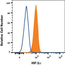
- Detection of HIF-1 alpha in MCF-7 Human Cell Line by Flow Cytometry. MCF-7 human breast cancer cell line treated with CoCl2 was stained with Mouse Anti-Human/Mouse HIF-1 alpha APC-conjugated Monoclonal Antibody (Catalog # IC1935A, filled histogram) or isotype control antibody (Catalog # IC002A, open histogram). To facilitate intracellular staining, cells were fixed with Flow Cytometry Fixation Buffer (Catalog # FC004) and permeabilized with Flow Cytometry Permeabilization/Wash Buffer I (Catalog # FC005). View our protocol for Staining Intracellular Molecules.
