 Explore
 Validate
 Learn
LearnGTX30115
antibody from GeneTex
Targeting: HIF1A
bHLHe78, HIF-1alpha, HIF1, MOP1, PASD8
 Western blot
 ELISA Immunocytochemistry Immunoprecipitation Immunohistochemistry Flow cytometry Gel shift Chromatin Immunoprecipitation
Antibody data
- Antibody Data
- Antigen structure
- References [2]
- Comments [0]
- Validations
- Western blot [2]
- Immunocytochemistry [1]
- Immunoprecipitation [1]
- Immunohistochemistry [3]
Submit
Validation data
Reference
Comment
Report error
- Product number
- GTX30115 - Provider product page

- Provider
- GeneTex
- Proper citation
- GeneTex Cat#GTX30115, RRID:AB_368095
- Product name
- HIF1 alpha antibody [H1alpha67]
- Antibody type
- Monoclonal
- Reactivity
- Human, Mouse, Rat, Bovine, Canine, Chicken/Avian, Porcine, Sheep, Simian
- Host
- Mouse
Submitted references Hypoxia-induced abrogation of contact-dependent inhibition of rheumatoid arthritis synovial fibroblast proliferation.
Hypoxia-inducible factor-1alpha and hypoxia-inducible factor-2alpha are expressed in kaposi sarcoma and modulated by insulin-like growth factor-I.
Nonomura Y, Mizoguchi F, Suzuki A, Nanki T, Kato H, Miyasaka N, Kohsaka H
The Journal of rheumatology 2009 Apr;36(4):698-705
The Journal of rheumatology 2009 Apr;36(4):698-705
Hypoxia-inducible factor-1alpha and hypoxia-inducible factor-2alpha are expressed in kaposi sarcoma and modulated by insulin-like growth factor-I.
Catrina SB, Botusan IR, Rantanen A, Catrina AI, Pyakurel P, Savu O, Axelson M, Biberfeld P, Poellinger L, Brismar K
Clinical cancer research : an official journal of the American Association for Cancer Research 2006 Aug 1;12(15):4506-14
Clinical cancer research : an official journal of the American Association for Cancer Research 2006 Aug 1;12(15):4506-14
No comments: Submit comment
Supportive validation
- Submitted by
- GeneTex (provider)
- Main image

- Experimental details
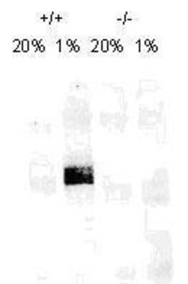
- Western Blot: HIF-1 alpha Antibody (H1alpha67) (GTX30115) - On day 1, MEF cells (+/+,-/-), were grown on 15 cm dish (2x10 to the 6th cells). On day 2, cells were exposed to hypoxia for 4hrs. Cells were washed with ice cold PBS twice and whole cell protein was extracted with RIPA buffer fortified with protease. Upon quantification, 100μg of protein was fractionated on 7% polyacralymide gel. Gel was transferred overnight onto nitrocellulose membrane. The membrane was probed with HIF-1 alpha monoclonal antibody at a 1:500 dilution (GTX30115). The secondary antibody was conjμgated with HRP and was used at a 1:2500 dilution
- Submitted by
- GeneTex (provider)
- Main image

- Experimental details
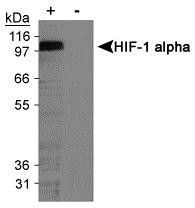
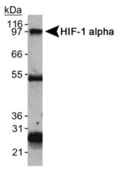
- WB analysis of cobalt chloride treated and untreated COS-7 nuclear extracts using HIF1 alpha antibody [H1alpha67].
Supportive validation
- Submitted by
- GeneTex (provider)
- Main image
- Experimental details
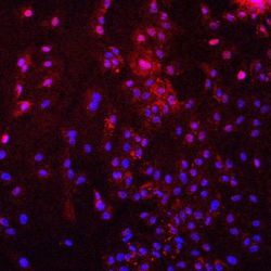
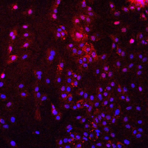
- ICC/IF analysis of pig endothelial cells under hipoxia condition using HIF1 alpha antibody [H1alpha67] (red) and DAPI.
Supportive validation
- Submitted by
- GeneTex (provider)
- Main image
- Experimental details
- Immunoprecipitation of HIF1 alpha protein using HIF1 alpha mouse monoclonal antibody [H1alpha67]. The heavy and light chains are also detected.

Supportive validation
- Submitted by
- GeneTex (provider)
- Main image
- Experimental details
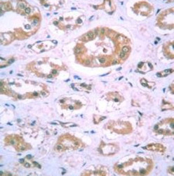
- Immunohistochemical staining of human kidney tissue using HIF1 alpha mouse monoclonal antibody [H1alpha67].

- Submitted by
- GeneTex (provider)
- Main image
- Experimental details
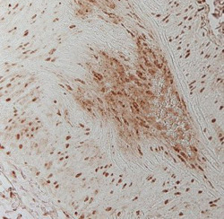
- Immunohistochemical staining of human lung tissue using HIF1 alpha mouse monoclonal antibody [H1alpha67].

- Submitted by
- GeneTex (provider)
- Main image
- Experimental details
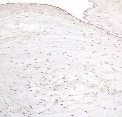
- IHC-P analysis of pig tissue (brown) using HIF1 alpha antibody [H1alpha67].
