 Explore
 Validate
 Learn
Learn Western blot
Antibody data
- Antibody Data
- Antigen structure
- References [1]
- Comments [0]
- Validations
- Western blot [1]
- Immunocytochemistry [1]
- Immunohistochemistry [1]
Submit
Validation data
Reference
Comment
Report error
- Product number
- TA324790 - Provider product page

- Provider
- OriGene
- Product name
- Rabbit polyclonal HIF1A Antibody (N-term)
- Antibody type
- Polyclonal
- Description
- Rabbit polyclonal HIF1A Antibody (N-term)
- Host
- Rabbit
- Conjugate
- Unconjugated
- Epitope
- HIF1A
- Isotype
- IgG
- Antibody clone number
- NULL
- Vial size
- 400 µl
- Concentration
- 2.0 mg/ml
Submitted references Inhibition of Connexin 43 translocation on mitochondria accelerates CoCl2-induced apoptotic response in a chemical model of hypoxia.
Pecoraro M, Pinto A, Popolo A
Toxicology in vitro : an international journal published in association with BIBRA 2018 Mar;47:120-128
Toxicology in vitro : an international journal published in association with BIBRA 2018 Mar;47:120-128
No comments: Submit comment
Supportive validation
- Submitted by
- OriGene (provider)
- Main image

- Experimental details
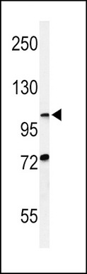
- Western blot analysis of HIF1A Antibody (N-term) (Cat. #TA324790) in CHO cell line lysates (35ug/lane). HIF1A (arrow) was detected using the purified Pab.
- Validation comment
- WB
Supportive validation
- Submitted by
- OriGene (provider)
- Main image

- Experimental details
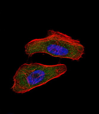
- IF image of Hela cell stained with HIF1A Antibody (N-term)(Cat#TA324790). Hela cells were incubated with HIF1A primary antibody (1:25, 1 h at 37?). For secondary antibody, Alexa Fluor? 488 conjugated donkey anti-rabbit antibody (green) was used (1:400).Cytoplasmic actin was counterstained with Alexa Fluor? 555 (red) conjugated Phalloidin (7 units/ml). Nuclei were counterstained with DAPI (blue) .HIF1A immunoreactivity is localized to cytoplasm and nucleus significantly.
- Validation comment
- IF
Supportive validation
- Submitted by
- OriGene (provider)
- Main image
- Experimental details
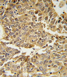
- HIF1A Antibody (N-term)(Cat. #TA324790) IHC analysis in formalin fixed and paraffin embedded lung carcinoma followed by peroxidase conjugation of the secondary antibody and DAB staining. This data demonstrates the use of the HIF1A Antibody (N-term) for immunohistochemistry. Clinical relevance has not been evaluated.
- Validation comment
- IHC
